
Interactive guide

Your spine MRI report, decoded.

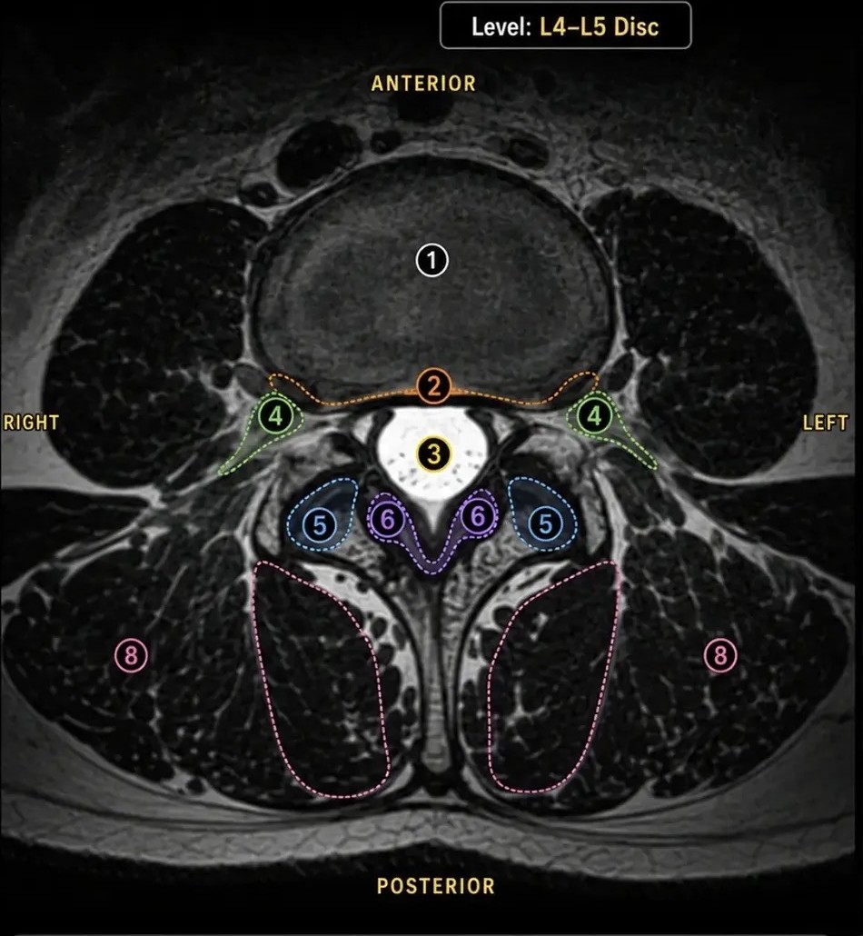
Below is a realistic sample MRI report, the kind of language you might see in your own results. Click each numbered finding to read what the report says, and then what it actually means. Then zoom into a labeled neck or low-back scan, one structure at a time.

Medically reviewed by Barrett S. Boody, M.D. · Updated September 2026

Go to an emergency department if you have a new change in bowel or bladder control, numbness in the groin or inner thighs, or weakness that is getting worse over hours or days. Those findings need same-day evaluation, not a website.

Interactive sample report

The click-through viewer needs JavaScript. Every finding in the sample report is written out further down this page.

What does the “Impression” mean?

Every MRI report ends with an Impression, the radiologist's summary of what matters. It is the part most patients read first and understand least. Here is this sample's impression, line by line.

Disc degenerative changes in the cervical and lumbar spine.

Normal age-related wear of the discs in the neck and lower back. It is the most common finding on adult spine MRIs and, on its own, not a diagnosis.

Posterior disc bulge at C5-C6 with mild canal stenosis.

A neck disc pressing back on the sac that holds the spinal cord, with mild narrowing of the canal. Mild, and the cord looks healthy. See this level on a labeled cervical MRI.

Small central disc protrusion at T7-T8 without significant stenosis.

A small mid-back disc finding that is not narrowing the canal. Typically not a source of symptoms.

Broad-based disc bulge at L4-L5 with mild-to-moderate canal stenosis and mild foraminal narrowing.

The most significant finding here: lower-back narrowing that can cause leg symptoms with standing or walking, if it lines up with how you feel. Lumbar spinal stenosis.

Disc bulge at L5-S1 touching the S1 nerve roots.

The lowest disc resting against the sciatic nerve roots. A level worth watching if you have leg pain. Sciatica and herniated disc.

Sacroiliac joints, sacrum, and coccyx are normal.

No problem found at the SI joints or tailbone. Reassuring and normal.

Read all five sample findings

The body of the report walks down the spine from the neck to the tailbone, one level at a time. Each finding below pairs the radiologist's sentence with a plain-English translation. This is a sample for education, not your scan.

Cervical spine, C5 to C6

Posterior disc bulge at C5-C6 indenting the anterior thecal sac. Mild spinal canal stenosis. No significant neural foraminal narrowing. Cervical cord signal is normal.

In plain English: The cushion between the 5th and 6th neck vertebrae is spreading backward and gently pressing on the front of the sac that holds your spinal cord, slightly narrowing the canal. The narrowing is mild, the side doorways where nerves exit are open, and the spinal cord itself looks healthy. This is a very common neck finding.

See this level on a labeled cervical MRI →

Thoracic spine, T7 to T8

Small central disc protrusion at T7-T8 mildly indenting the anterior thecal sac. No significant spinal canal stenosis. No neural foraminal narrowing. Thoracic cord signal is normal.

In plain English: In the mid-back, a small focal bit of disc is pushing straight backward and just touching the front of the nerve sac. There is no meaningful narrowing of the canal or the nerve exits, and the spinal cord looks normal. Mid-back disc findings like this are usually quiet and rarely cause symptoms.

Lumbar spine, L4 to L5

Broad-based posterior disc bulge at L4-L5 with mild ligamentum flavum hypertrophy. Mild to moderate central canal stenosis. Mild bilateral neural foraminal narrowing. No significant spondylolisthesis.

In plain English: In the lower back, the disc has spread evenly backward and the ligament lining the back of the canal has thickened a little. Together they narrow the main canal to a mild-to-moderate degree and slightly tighten the nerve exits on both sides. The vertebrae are still lined up (no slippage). This combination is the classic picture of spinal stenosis.

Lumbar spinal stenosis →

Lumbosacral junction, L5 to S1

Posterior disc bulge at L5-S1 abutting the bilateral traversing S1 nerve roots. Mild bilateral neural foraminal narrowing. No spondylolisthesis.

In plain English: At the very bottom of the spine, the disc is bulging back far enough to touch the S1 nerve roots on both sides as they pass by, without clearly compressing them. The nerve exits are mildly tight. The S1 nerve is the one that runs down the back of the leg, so this is a level that can produce sciatica when it is irritated.

Sciatica and herniated disc →

Sacrum and coccyx, Pelvis

Sacroiliac joints appear normal. No acute fracture or marrow edema. Coccyx is normal.

In plain English: The joints where the spine meets the pelvis (the SI joints), the bone itself, and the tailbone all look normal: no fracture, no sign of recent injury or inflammation. A clean bill of health for the bottom of the spine.

Zoom into one level
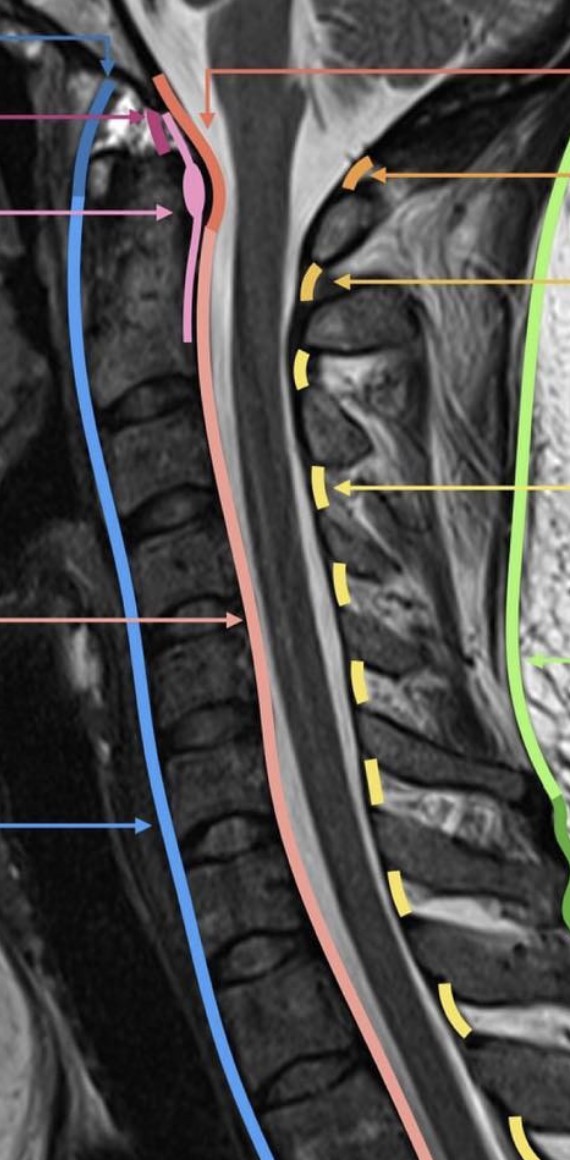
The report names structures: the thecal sac, the foramina, the ligamentum flavum, the facet joints. These two guides let you click each one on a real labeled scan and see what it is and why it matters.

The thoracic (mid-back) and sacral findings in this report are mild and are fully explained above.

How Dr. Boody reads it

The MRI is read next to the exam, never on its own.

Many adults with no symptoms have disc bulges, narrowing, and degenerative changes on a spine MRI. So the first question Dr. Boody asks of a report is not what it lists but which findings explain the symptoms and the exam in front of him. A level that matches is a target. A level that does not is noted and left alone.

In clinic he pulls up your images, not just the report, and marks the target on the screen. That is the same walkthrough his fellows at Indiana Spine Group get before a case, and it is why the explanation you leave with names the level, the structure, and the reason.

Some of the hardest calls live on the MRI. He has published on one of them in Clinical Spine Surgery: whether a person whose scan shows spinal cord compression with signal change, but who has no symptoms, should have surgery. When your report raises a question like that, the answer rests on published evidence and on your exam, and he will walk you through both.

Research and teaching

Questions patients ask

MRI-report jargon, translated.

What does “posterior disc bulge indenting the anterior thecal sac” mean?
It means a spinal disc has spread backward (posterior) and is gently pressing on the front (anterior) of the thecal sac, the fluid-filled sleeve that protects your spinal cord and nerves. Indenting means it is pushing in on the sac without necessarily pinching a nerve. It is one of the most common findings on a spine MRI and is often a normal age-related change that causes no symptoms.
What is central canal stenosis on an MRI?
Central canal stenosis means the main channel that carries the spinal cord and nerves has narrowed. Reports grade it as mild, moderate, or severe. Mild stenosis frequently causes no symptoms. Moderate to severe narrowing can cause pain, numbness, or heaviness, especially with standing or walking. Stenosis is interpreted alongside your symptoms, not in isolation.
What does “neural foraminal narrowing” mean?
The neural foramina are the small side doorways where a nerve root exits the spine on its way to the arm or leg. Foraminal narrowing means one of those doorways is tighter than normal, which can pinch the exiting nerve and cause radiating pain, tingling, or weakness. That pattern is called radiculopathy; in the leg it is often called sciatica.
What does “disc bulge abutting the traversing nerve roots” mean?
Abutting means the disc is touching or resting against the nerve roots without clearly compressing them. The traversing nerve roots are the nerves passing through that level on their way to the next exit. Whether this matters depends on your symptoms. Contact between a disc and a nerve root is common and does not always cause pain.
Is it serious to have disc bulges at several levels of my spine?
Usually not. Disc bulges and degenerative changes at multiple levels are extremely common as we age and are found on the MRIs of many people who have no pain at all. What matters is whether a finding lines up with your specific symptoms and physical exam. Dr. Boody correlates the imaging with how you feel and what the exam shows to decide what, if anything, needs treatment.
What is the difference between a disc bulge and a disc protrusion?
A bulge is a broad, even spreading of the disc beyond its normal edge, like a tire that sags outward all the way around. A protrusion is more focal: a smaller, localized push of disc material in one spot. Both are common. A protrusion is slightly more likely to contact a nerve because it is concentrated in one area.
What does “cord signal is normal” mean, and what if it is not?
On T2-weighted images the spinal cord should look an even grey. A bright spot inside it, called signal change, means the cord tissue itself has been affected by pressure, not just the space around it, and the term myelomalacia describes cord that has been damaged by long-standing compression. The sample report says the cord signal is normal at every level. When it is not, the finding is weighed with the exam. Dr. Boody has published on whether cord compression with signal change but no symptoms should be treated.
Is this my MRI?
No. The report on this page is a realistic sample used for education only. It is not your personal scan. Dr. Boody reviews your own imaging with you during your consultation and explains exactly what it shows for your specific situation.

Next step

Have a report of your own?

Bring it, and bring the images. Dr. Boody walks through your actual scan with you, finding by finding, and you leave knowing which ones matter and which do not. Offices in Carmel, Fishers, Greenfield, and Muncie.