Treatments

Non-surgical first. Then the smallest operation that reliably works.

Dr. Boody's practice covers the full range, from injections to complex revision and deformity surgery. That range is the point: when every option is on the table, the recommendation can be the one that fits your problem rather than the one a surgeon happens to offer. Each treatment below explains what it does, what it is for, and what it cannot fix.

Non-surgical first

Most spine problems never need an operation.

Disc herniations shrink, inflamed nerves calm down, and strained backs heal. The job of the first weeks is to control pain and protect function while that happens. Surgery is for the problems that do not resolve, and for a short list that should not wait.

Physical therapy

Targeted exercise to take load off the painful structure, restore motion, and build the strength that keeps the problem from returning. For nerve pain, therapy is about positions and movement that give the nerve room. It is the foundation of non-surgical care and of recovery after surgery.

Medication

Anti-inflammatories for pain from an irritated disc or joint, nerve-pain medications for radiating symptoms, and a short course of oral steroids in some acute cases. Medication treats the pain while the body heals; it does not change what the MRI shows, and long-term opioids are not a treatment for spine pain.

Injections

Epidural steroid injections put anti-inflammatory medication next to an irritated nerve root. Facet and sacroiliac joint injections treat, and help diagnose, pain from a specific joint. Relief may last weeks to months, which can be enough for the underlying problem to settle, and a good response confirms which structure is responsible.


When non-surgical care fits

  • Neck or back pain without nerve symptoms
  • Arm or leg pain from a pinched nerve with no significant weakness
  • Stenosis symptoms you can still walk through
  • A stable fracture that can heal in a brace
  • Any problem where the MRI finding does not clearly explain the symptoms

When it does not, and surgery should be discussed now

  • Weakness that is progressing
  • Cervical myelopathy: clumsy hands, unsteady walking, spinal cord compression on MRI
  • An unstable fracture or an injury with any nerve or cord involvement
  • Infection with an abscess pressing on nerves, or bone destruction
  • Bowel or bladder change or saddle numbness, which is an emergency

Surgical procedures

Neck operations.

Most cervical surgery is done from the front of the neck, through a small incision beside the windpipe. The disc pressing on the nerve or cord is removed, and the space is either fused or replaced with a moving implant. Which one is the decision Dr. Boody has studied longest.

Images are simplified teaching models. Each full guide takes a closer look.

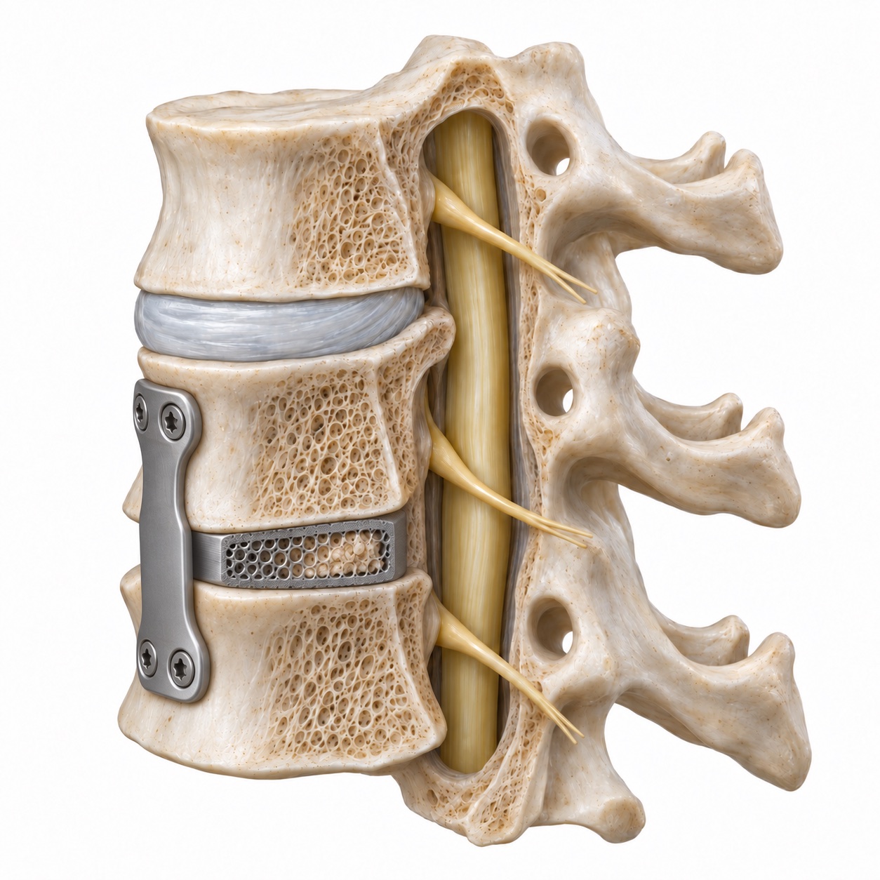
Cervical fusion model with a cage and anterior plate
Fusion support

ACDF (neck fusion)

Relieves nerve or spinal cord pressure, then joins the treated vertebrae together.

What it involves: ACDF (neck fusion)

The disc and any bone spurs pressing on the nerve or cord are removed from the front of the neck, and the space is filled with a bone graft or cage held by a small plate so the two vertebrae grow together. It relieves nerve and cord compression reliably and is the standard operation for cervical radiculopathy and myelopathy. The trade-off is that the fused level no longer moves, which places slightly more work on the levels next to it.

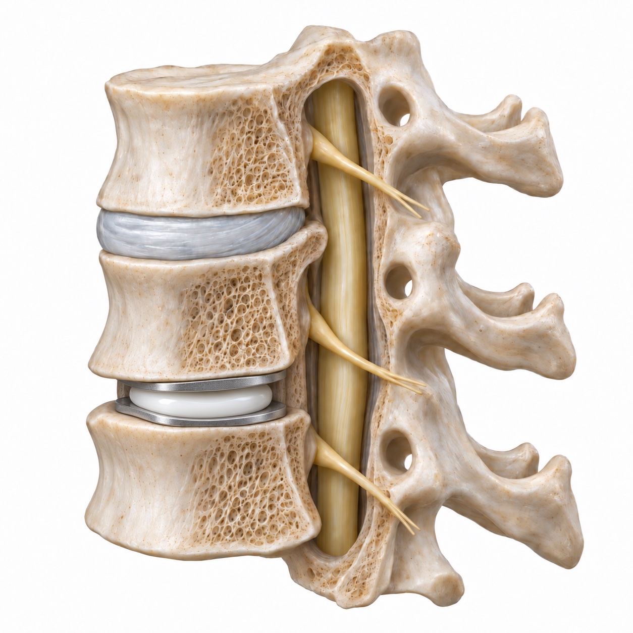
Cervical model with an artificial disc between two vertebrae
Moving implant

Cervical disc replacement

Relieves pressure with an artificial disc designed to preserve motion at the treated level.

What it involves: Cervical disc replacement

The same decompression as an ACDF, but the disc is replaced with an implant that lets the level keep moving instead of fusing it. The aim is to protect the neighboring levels from the extra load a fusion places on them. It suits patients with good bone, well-preserved facet joints, and disease at one or two levels; it is not the right choice for every neck. Dr. Boody co-authored twenty-year outcomes from a randomized trial of this operation.

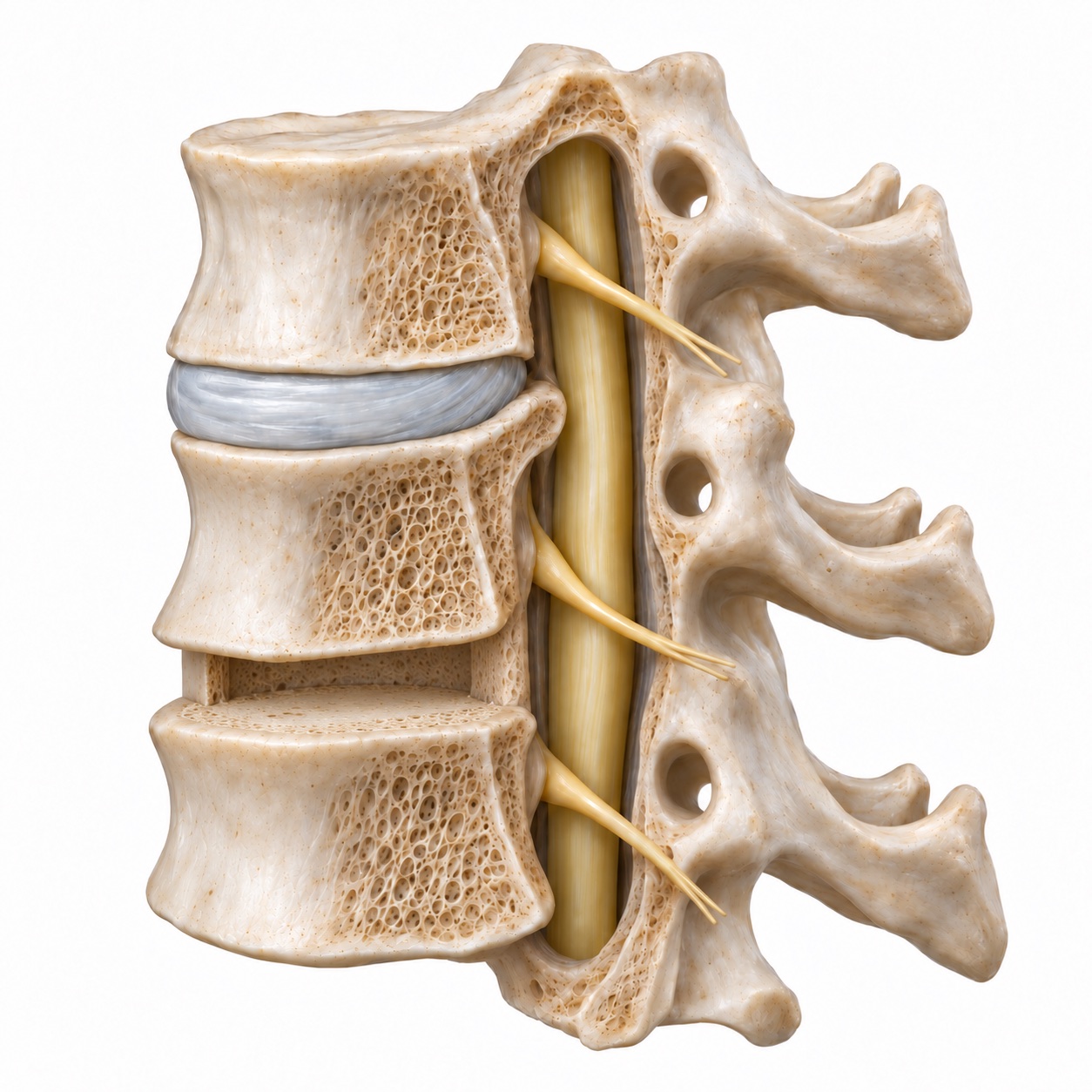
Cervical teaching model showing space after a disc is removed
Decompression example

Spinal cord decompression

Creates more room for the spinal cord, using the approach that fits the source of pressure.

What it involves: Spinal cord decompression

Any operation whose purpose is to take pressure off the spinal cord, in the neck or the mid-back. Depending on where the compression comes from and how many levels are involved, it is done from the front (removing discs or a vertebral body), from the back (laminectomy or laminoplasty, which opens or hinges the roof of the canal), or both. It is the treatment for myelopathy, OPLL, and cord compression from fracture or tumor. Its goal is to stop the cord from getting worse; recovery of function already lost is possible but not guaranteed.

Surgical procedures

Low back operations.

The lumbar question is usually whether the nerve needs to be freed, or whether the level also needs to be held still. Decompression alone is a smaller operation; fusion is added when the spine is unstable or slipping.

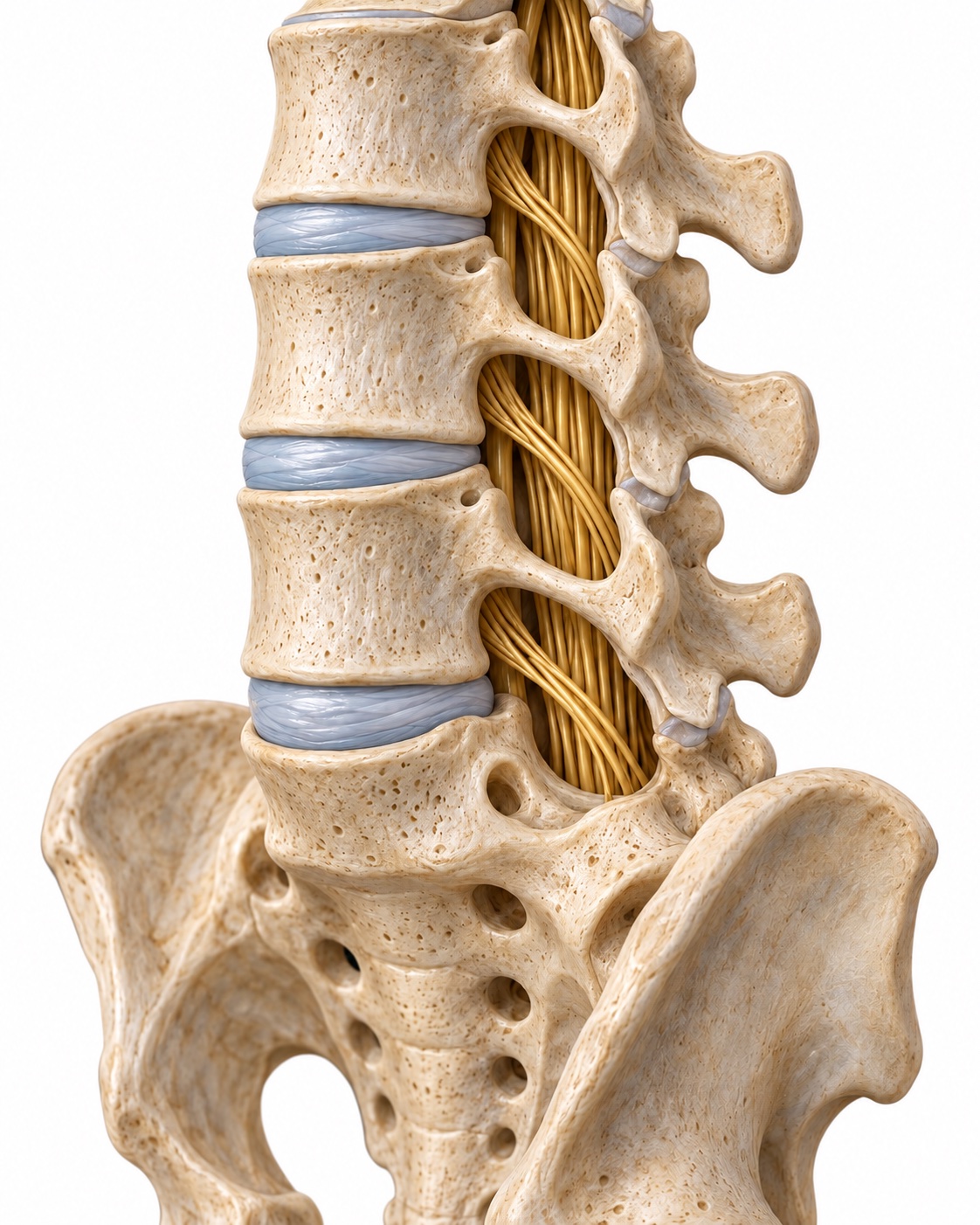
Anatomical model of the lumbar spine and exiting nerves
Lumbar anatomy

Far lateral microdiscectomy

Reaches a disc fragment outside the nerve tunnel through an approach from the side.

What it involves: Far lateral microdiscectomy

A microdiscectomy removes the fragment of disc pressing on a nerve root through a small incision, using a microscope. When the fragment sits out beyond the edge of the canal, where the nerve has already exited, a standard approach cannot reach it without removing part of the facet joint. The far lateral approach comes in from the side, reaches the nerve directly, and leaves the joint intact. Dr. Boody has published on the technique.

Lumbar fusion construct with a cage, screws and rods
Fusion support

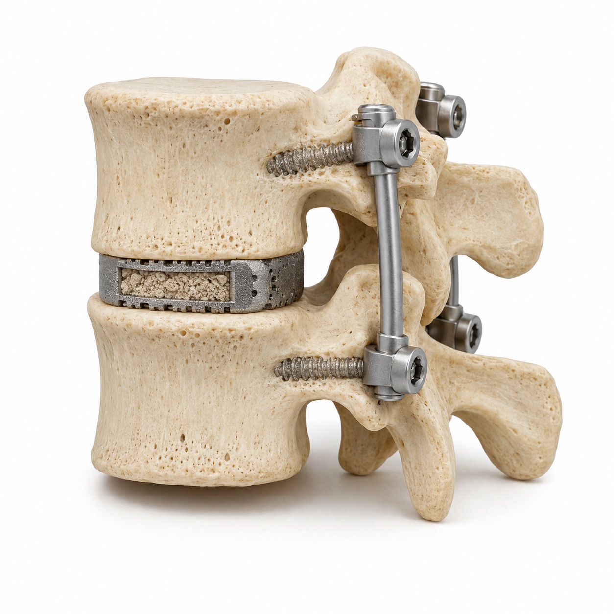
Lumbar fusion (TLIF)

Combines nerve decompression with support for a level that needs to heal together.

What it involves: Lumbar fusion (TLIF)

A fusion of one or more lumbar levels done from the back. The worn disc is removed through the opening the nerve exits from, a cage with bone graft is placed in the disc space to restore its height and alignment, and screws hold the level still while the bone heals into a single unit. It is the usual operation for spondylolisthesis and for stenosis at an unstable level, and it can be done through small incisions with tubular retractors.

Anatomical model of the lower spine and pelvis
Spine and pelvis

Sacroiliac joint injections

Targets the joint between the spine and pelvis to help evaluate and relieve pain.

What it involves: Sacroiliac joint injections

The sacroiliac joints connect the base of the spine to the pelvis, and they are a frequently missed source of low back and buttock pain, including after a lumbar fusion. An injection of anesthetic and steroid into the joint under X-ray guidance does two things: it relieves the pain for a period, and if it does, it confirms the joint as the source. That answer determines what comes next, and it is a reason not to operate on a disc that was never the problem.

Surgical procedures

Techniques, revision, and deformity.

Minimally invasive methods change how an operation is done, not what it accomplishes. Revision and deformity surgery are the other end of the range: larger operations for problems that a smaller one cannot fix.

Teaching model showing a narrow tube through back muscle toward the lumbar spine
Smaller working corridor

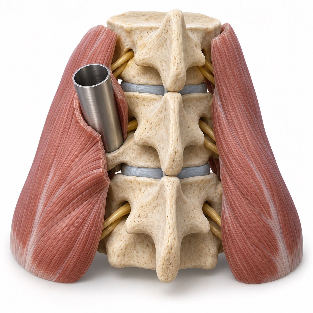
Minimally invasive spine surgery

Uses a smaller muscle corridor to carry out a decompression or fusion.

What it involves: Minimally invasive spine surgery

Doing the same decompression or fusion through small incisions, with tubular retractors that spread the muscle rather than cutting it, and X-ray or navigation guiding the instruments. Less muscle damage generally means less pain after surgery and a faster return to activity. It is a way of operating, not a separate operation, and it is used when it can accomplish the full goal; when it cannot, a larger incision is the safer choice.

Illustrative fusion hardware showing pedicle screws connected by rods
Fixation hardware example

Percutaneous pedicle screws

Places screws and rods through small incisions to support a fusion or stabilize a fracture.

What it involves: Percutaneous pedicle screws

Pedicle screws anchor a fusion or stabilize a fracture. Placing them percutaneously means each screw goes in through a small stab incision guided by X-ray or navigation, with rods threaded beneath the skin to connect them, instead of exposing the whole back of the spine. It is often used to stabilize a thoracolumbar fracture and as the fixation for a minimally invasive TLIF. Dr. Boody has studied how surgeons learn to place these screws safely.

Lumbar anatomical model with wear above a previous fusion
Previous fusion

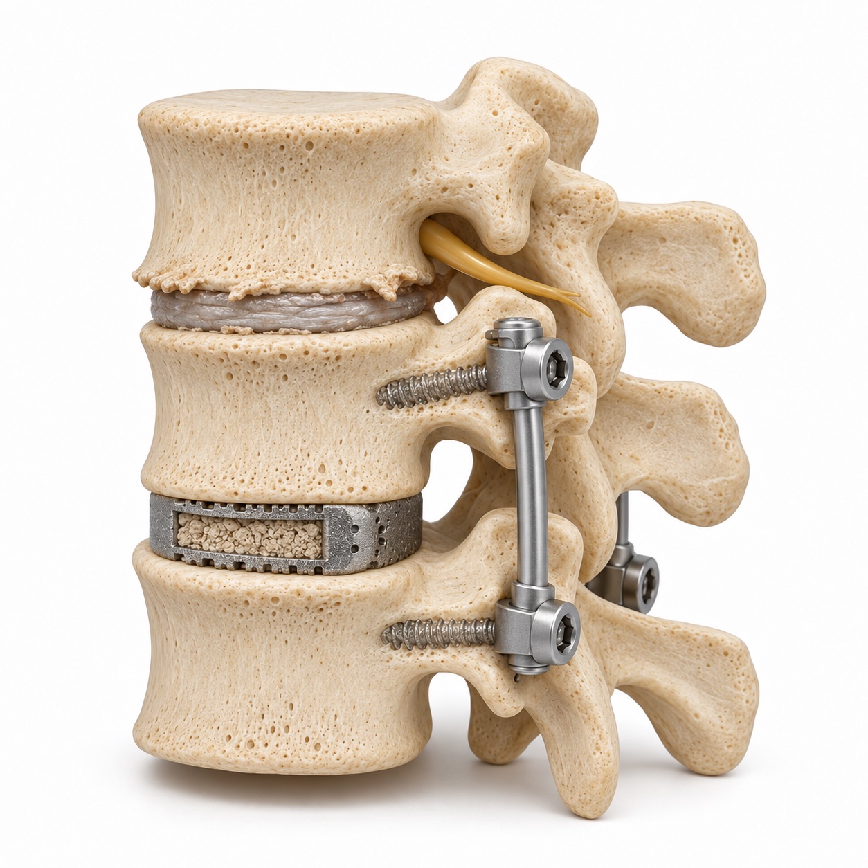
Revision spine surgery

Addresses a specific problem at or near a level that has already had surgery.

What it involves: Revision spine surgery

Surgery on a spine that has already been operated on: a fusion that did not heal, a level wearing out next to a fusion, hardware that has loosened or was placed in the wrong spot, or a problem the first operation did not fully address. Revision is harder than a first operation because of scar tissue and changed anatomy, and it starts with an honest diagnosis of why the first surgery did not work. Dr. Boody takes revision cases from his own practice and from surgery done elsewhere.

Deformity correction

Treats spinal curves or imbalance when the alignment itself is causing the problem.

What it involves: Deformity correction

Surgery to straighten a scoliosis curve or restore the side-view alignment that keeps the head balanced over the pelvis, using screws and rods across multiple levels, sometimes with bone cuts (osteotomies) to allow the correction. In adolescents it stops a large curve from progressing; in adults it treats pain and nerve symptoms caused by the alignment itself. It is the largest operation in spine surgery, and the decision to do it is made only after conservative care, and only when the deformity is clearly the cause.

Appointments

Find out which of these, if any, is right for you.

The visit does not assume an operation at the end of it. Bring your imaging and what you have already tried, and you will leave with the options ranked and explained.