

What is cervical disc replacement?
See the difference
After the damaged disc is removed.
Both operations relieve pressure on the nerves or spinal cord. Compare what goes into the disc space and what that means for motion.
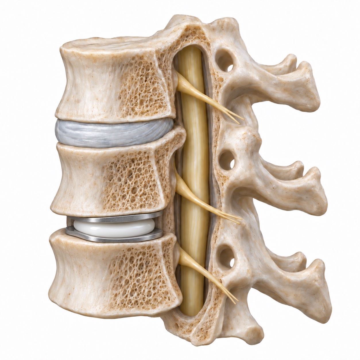
An implant designed to move
The artificial disc has surfaces that attach to the bones and a bearing that allows movement between them. The goal is to preserve motion at a level that still moves.
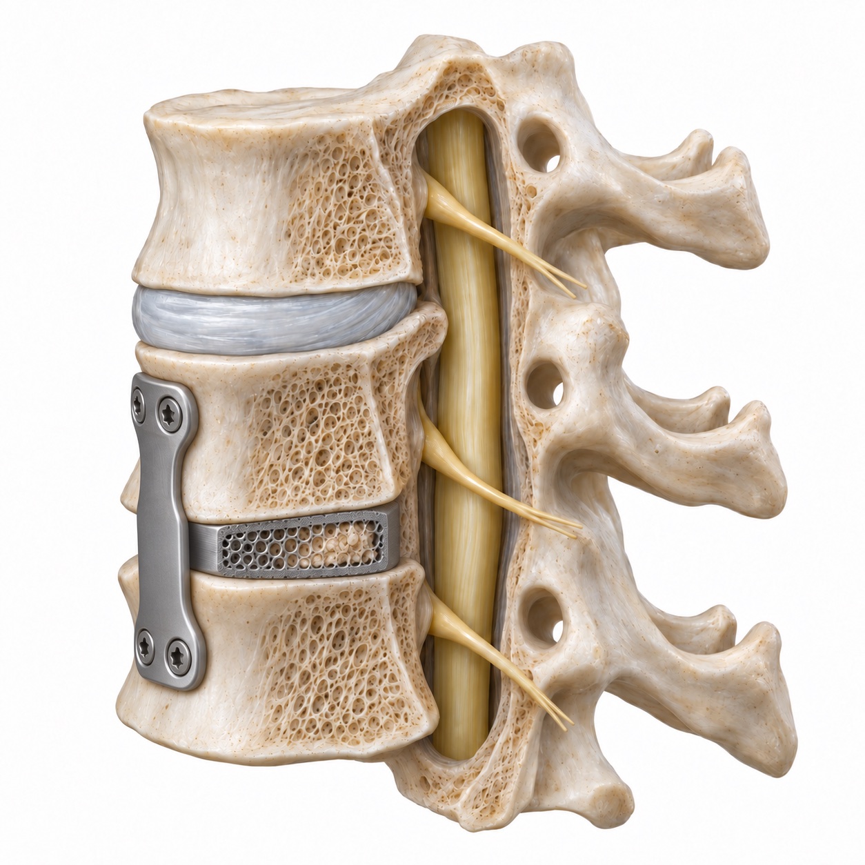
A level intended to heal together
A spacer with bone graft supports the disc space while the two vertebrae fuse. The goal is a stable bone connection rather than continued motion at that level.
Simplified anatomical models, not patient scans or specific implant recommendations. Preserving motion does not guarantee a better outcome for every patient.
More about artificial discs and motion preservation
A disc is the cushion between two vertebrae. When it herniates or wears down, it and the bone spurs that grow around it can press on a nerve root or the spinal cord. For decades the standard fix was to remove the disc and fuse the two bones together. Fusion works well, but it costs the motion at that level and shifts more work onto the levels above and below.
An artificial cervical disc is designed to keep that motion. The implant has two metal endplates that anchor to the vertebrae above and below, with a bearing surface between them, usually a durable plastic core or a metal-on-metal articulation depending on the design. The implant allows the level to flex, extend, bend sideways, and rotate within a controlled range, much like the disc it replaced. Several designs have been approved by the FDA for one-level use, and some for two adjacent levels.
The theory behind motion preservation is simple. If the treated level keeps moving, the neighboring levels are not asked to move more, so they should wear more slowly. Whether that theory holds up over the long term is a question that could only be answered by following patients for a long time. Dr. Boody has been part of answering it.
Who qualifies for disc replacement, and who does not?
Does the level still move?
The aim is to preserve useful motion. A level that is already stiff or collapsed may be better treated with fusion.
Are the joints behind it healthy?
Arthritis in the facet joints can make movement painful. Keeping that movement may not help.
Can the bone support the implant?
Bone quality and stability matter. Imaging helps determine whether replacement is appropriate.
Read the full eligibility criteria and reasons to consider fusion instead
Disc replacement is considered when you have cervical radiculopathy, a pinched nerve root causing arm pain, tingling, or weakness, or early cervical myelopathy from a single soft disc pressing on the cord, and non-surgical care has not solved it. The FDA-studied indications are one or two levels between C3 and C7 in adults with mature bone.
The level has to be able to move. An implant placed at a level that has already collapsed and stiffened has nothing to preserve, and it is more likely to be surrounded by new bone. The facet joints at the back of the level need to be healthy; arthritis there causes pain with motion, which is the last thing you want to keep. There must be no instability, meaning the vertebrae do not slip on each other on bending X-rays.
Reasons disc replacement is usually not recommended include significant facet arthritis, instability or a prior fusion at the same level, osteoporosis, long-segment compression of the spinal cord, ossification of the posterior longitudinal ligament (OPLL), active infection, and rheumatoid disease of the neck. In those settings fusion is the more reliable operation. Dr. Boody will look at your MRI, standing X-rays, and bending X-rays before telling you which group you fall into.
What happens during the operation?
Through the operation
From pressure to replacement.
Step through the same anatomical view. The lower disc space is the treated level in each illustration.
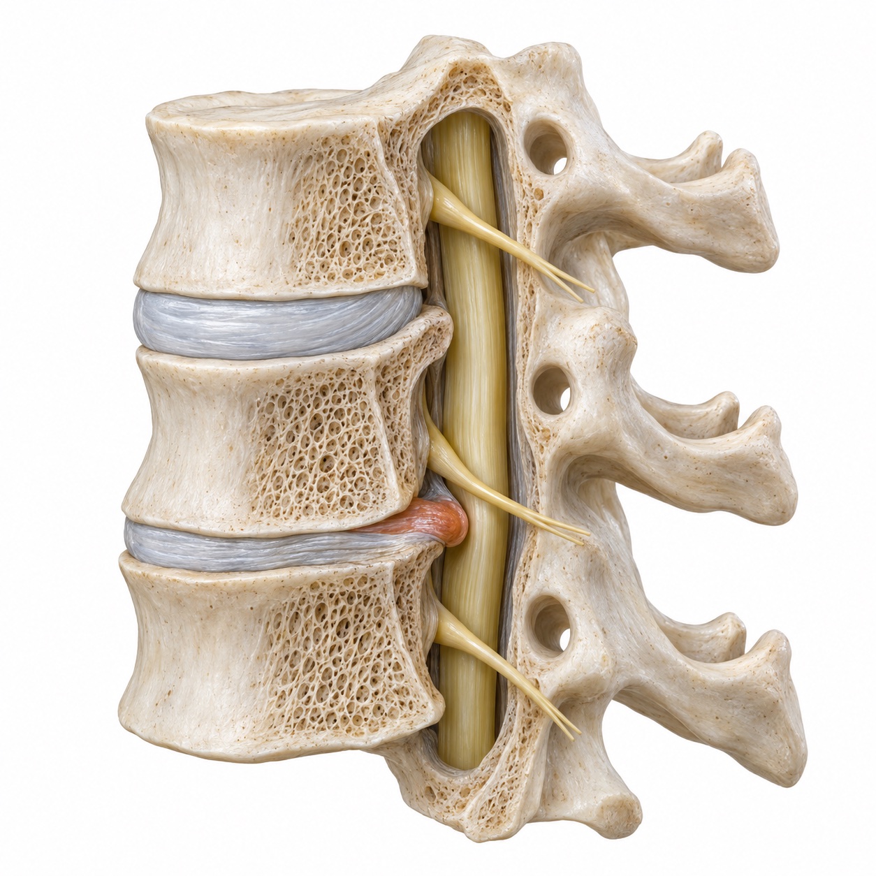
Find the source of pressure
The disc bulge shown in terracotta crowds the space beside the spinal cord. The examination and imaging identify which level is responsible for the symptoms.
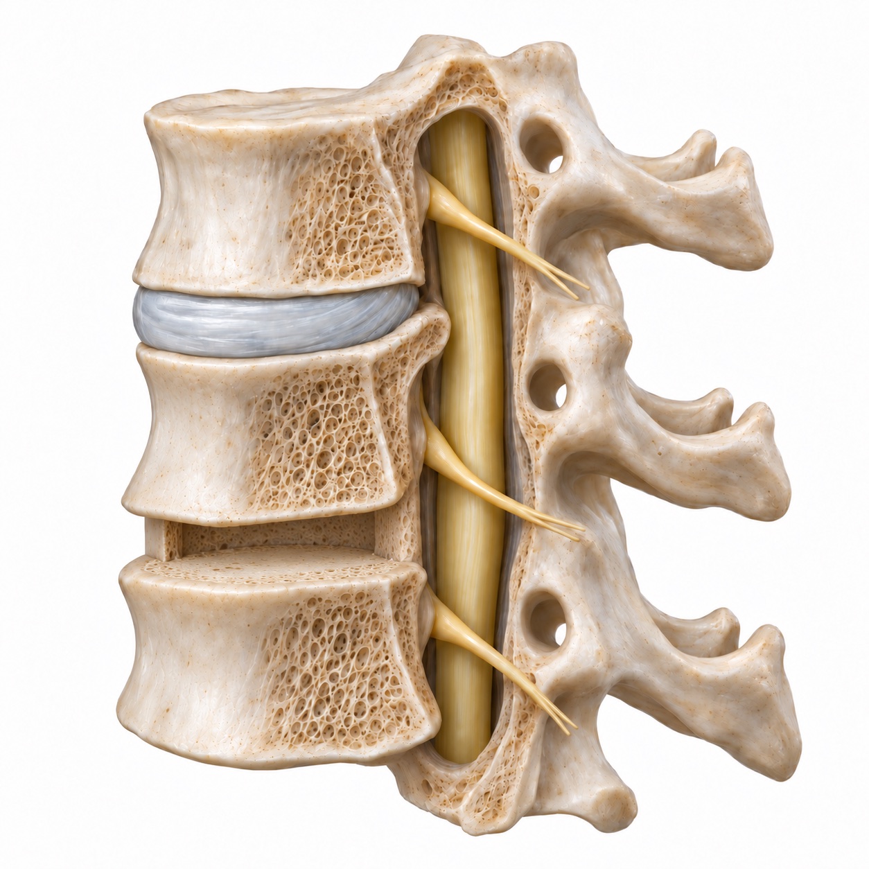
Remove the disc and free the nerves
Through the front of the neck, the surgeon removes the damaged disc and any relevant bone spurs to relieve pressure. The bone surfaces are prepared for the implant.
Fit the artificial disc
An implant is selected to fit the level and seated between the vertebrae. X-rays help check its size and position.
Illustrations omit surrounding soft tissue and instruments. The implant shown is a generic teaching model; the device and surgical plan depend on your anatomy.
Read the full surgical steps, from anesthesia to implant positioning
The first two thirds of the operation are identical to ACDF, and that is by design: the goal of decompressing the nerve is the same, only the ending differs.
- Anesthesia. You are fully asleep with a breathing tube and monitored by an anesthesiologist. Nerve monitoring is used when the spinal cord is involved.
- Incision. A horizontal incision about two inches long in a skin crease on the front of the neck, usually on the left.
- Reaching the spine. The surgeon passes between the windpipe and swallowing tube on one side and the carotid artery and jugular vein on the other, without cutting major muscle. An X-ray confirms the level.
- Removing the disc. Under magnification, the disc and bone spurs are removed and the nerve tunnels are opened. Compared with a fusion, the surgeon is more careful to keep the bony endplates flat and intact, because the implant has to sit on them.
- Sizing and placing the implant. Trial implants are used to find the right height and footprint. The implant is then set in the middle of the disc space under X-ray guidance. Its position matters for how well the level moves afterward.
One level typically takes one to two hours. A two-level replacement adds roughly forty-five minutes to an hour. Most patients are discharged the same day or the next morning.
What are the alternatives?
ACDF. The same approach and the same decompression, followed by a fusion instead of an implant. It works at levels that are too stiff, arthritic, or unstable for a replacement, and it has the longest track record. Its costs are the lost motion at the level and the extra load on the neighbors over time.
Posterior foraminotomy. When a herniation is off to the side and pinches only the exiting nerve root, a small window made from the back can free it without any implant. It does not address the center of the canal or restore disc height.
Use the visual comparison above to see the two approaches, or read more on the ACDF page. The short version: disc replacement keeps motion and, in long-term trials, has been associated with less degeneration at the neighboring levels; ACDF is the more forgiving operation when the level is already worn.
What are the risks?
- Swallowing soreness and hoarseness. Common for one to two weeks because the swallowing tube is moved aside; usually resolved within six weeks. Temporary hoarseness in a minority of patients; permanent voice change is rare.
- Heterotopic ossification. This is the honest trade-off. In some patients, bone gradually forms around the implant. Studies report it in a substantial share of patients over the years, with wide variation depending on the device and how it is graded. In most it causes no symptoms. In some it reduces the motion the implant was placed to preserve, and in a minority the level eventually stiffens into what amounts to a fusion. Anti-inflammatory medication in the weeks after surgery is often used to lower the risk.
- Implant position or movement. An implant that is placed off-center or that shifts can move poorly or irritate nearby structures. This is uncommon and is the reason the operation is done under repeated X-ray checks. Subsidence, where the implant settles into softer bone, is another reason osteoporosis rules the procedure out.
- Conversion to fusion. A small share of patients need a later operation at the same level, most often for persistent pain from the facet joints or for an implant problem. That operation is a fusion.
- Nerve or spinal cord injury, bleeding, infection, blood clots, anesthesia. All uncommon, and the same as for any anterior neck operation.
What is recovery like?
Most people go home the same day or after one night, then build activity gradually.
There is no fusion to wait for, but the incision and tissues still need time to heal.
- First week
Walk and ease into gentle movement.
A sore throat and neck stiffness are common early on. Short walks and gentle neck motion accompany limits on lifting.
- Weeks 2 to 6
Return to your daily routine.
Swallowing improves and walking increases. Driving waits until you are off narcotic medication and can turn comfortably.
- Months 2 to 3
Build strength and comfortable motion.
Therapy may help with posture and range of motion. Work and exercise increase gradually as recovery allows.
- Months 3 to 12
Keep an eye on the implant.
Follow-up imaging checks the implant and neighboring levels. Sport and heavier activity require clearance based on healing.
More about activity limits and recovery timing
First week
Sore throat and neck stiffness. Soft foods. Walk several times a day. Gentle neck motion is encouraged rather than restricted. No lifting over about ten pounds. Pain medication for a few days; many patients are given a short course of anti-inflammatory medication to lower the chance of bone forming around the implant.
Weeks 2 to 6
Swallowing settles. Driving once off narcotic medication and turning comfortably. Desk work usually resumes by week two or three. Walking builds up; no overhead loading or impact yet. An X-ray checks implant position.
Months 2 to 3
Physical therapy for range of motion and posture if needed. Return to physical work and most exercise with a gradual ramp. Arm symptoms have usually improved substantially by now.
Months 3 to 12
Bone grows onto the implant endplates and anchors them. Full activity, including sport, is cleared once healing is confirmed. Periodic X-rays over the years watch implant motion and the neighboring levels.
What results can you expect?
The key word is selected. The trial results apply to patients who met the criteria described above. An implant placed at a stiff, arthritic level does not produce the same numbers, which is why candidacy is a decision made from your imaging rather than from your preference.
Dr. Boody co-authored the twenty-year radiographic results of a prospective randomized trial of single-level cervical disc arthroplasty, published in Spine in 2024. That is among the longest follow-up available for any spinal implant. The findings, in plain terms: the replaced levels continued to move two decades later, and the neighboring levels showed less degeneration than after fusion. Twenty years of data do not promise a specific outcome for you, but they answer the question patients most often ask, which is whether the motion lasts.
He also studied a subtler issue. In a 2020 paper in Clinical Spine Surgery, he examined adjacent-level ossification, the formation of bone at the levels next to the operated one, ten years after either disc replacement or ACDF, and how it related to patients' function. And in a 2019 paper in the International Journal of Spine Surgery, he and a colleague compared two-level ACDF with two-level arthroplasty, the setting where the choice between the two operations has the most long-term consequence, because two fused levels put more load on the neighbors than one.
The neck pain caveat applies here as it does to fusion: these operations treat arm symptoms and spinal cord compression reliably, and neck pain less reliably. If neck pain is your main complaint, expect a frank conversation about what the operation will and will not change.