

What is adjacent segment disease?
Look above the fusion
Same fusion. A different neighboring disc.
Switch between two illustrative examples. The cage and rods mark the lower treated level; the natural disc above it is the neighbor.
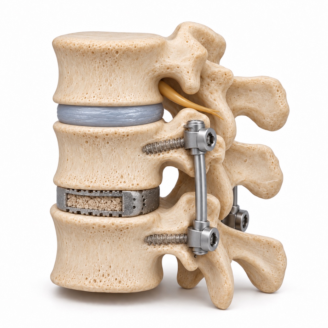
The adjacent level still has its own disc
The lower level is supported by a cage, screws, and rods. Above it, the natural disc remains a separate moving level.
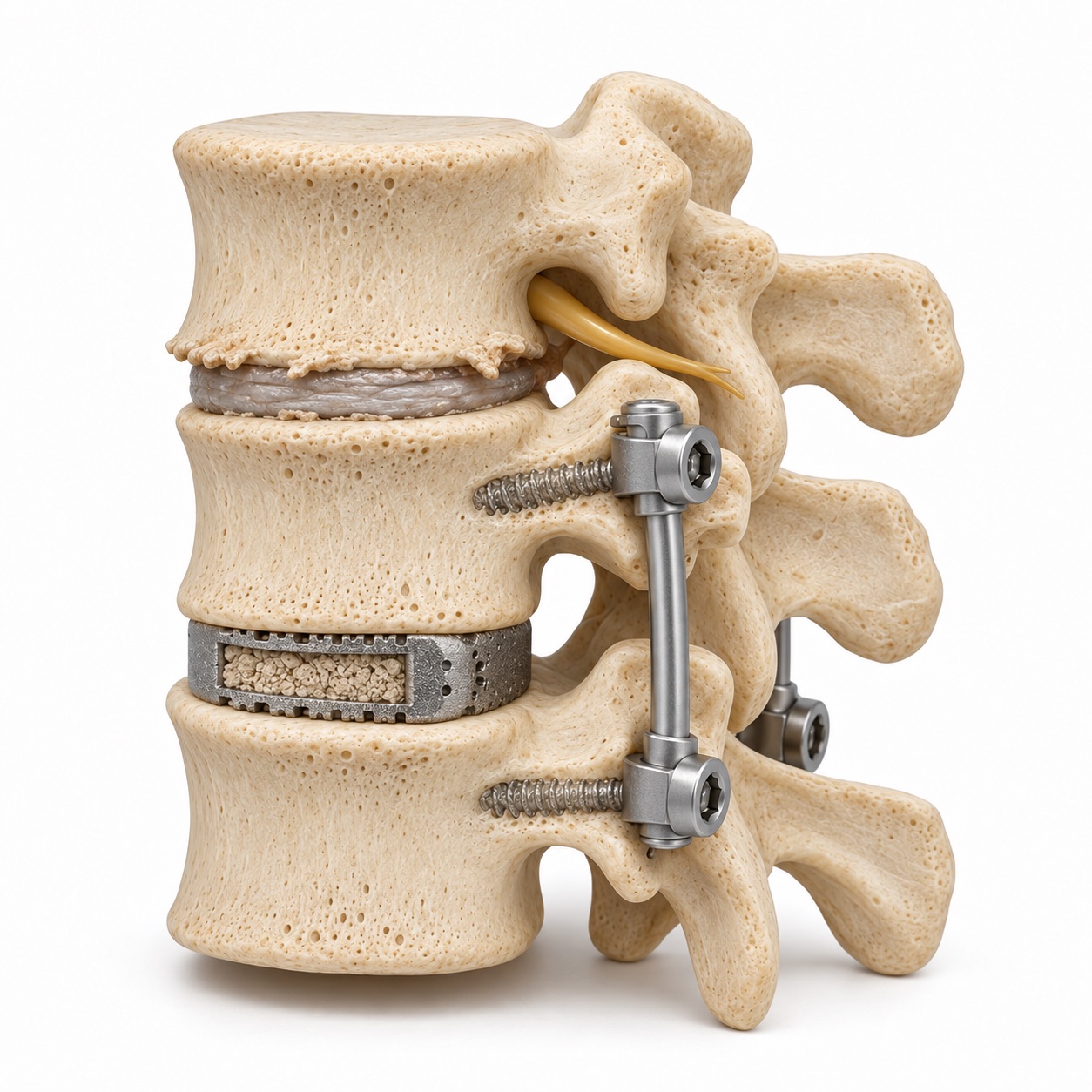
Wear develops at the neighboring level
The upper disc is thinner and its bone margins have developed spurs. These changes may narrow the space available for nerves.
These are simplified comparison models, not a patient’s before-and-after images or an inevitable sequence. Adjacent-level problems can occur in the neck as well as the low back.
Degeneration
Changes seen on imaging, which may cause no symptoms.
Symptomatic disease
New symptoms that correspond to the changes at the neighboring level.
Read more about the neighboring level and the meaning of imaging changes
A fusion is a deliberate trade. You give up motion at one or more levels to remove pain, stabilize a slip, or protect a nerve or the cord. In return, the spine has one less shock absorber, and the segments on either side of the fused block become the hinge points for movements that used to be shared. Over years, that extra work shows up on imaging as disc narrowing, bone spurs, and facet enlargement at the neighboring level. When those changes are found only on a scan, they are called adjacent segment degeneration. When they produce symptoms that bring you back to a spine surgeon, it is adjacent segment disease.
The distinction matters. Radiographic degeneration next to a fusion is common and usually silent. Symptomatic disease is less common, and it is the only version that needs treatment.
What causes it?
How load, alignment, and natural aging contribute
Biomechanical studies show that intradiscal pressure and motion at the level next to a fusion rise measurably compared with the same level in an unfused spine. The longer the fused block, the larger the lever arm and the greater the stress on the neighbor. A single-level fusion adds modest load; a multilevel fusion to the sacrum adds a great deal, which is why the level above a long lumbar construct is watched so carefully.
The first surgery can contribute in ways that have nothing to do with fusion itself. Pedicle screws placed too close to the facet joint above the fusion can damage that joint. Extensive stripping of the muscles and ligaments at the adjacent level takes away some of its natural support. A fusion left in a flattened or kyphotic position forces the neighbor to bend harder to keep your head over your pelvis. Each of these is avoidable, and avoiding them is part of doing the first operation well.
Finally, the disc next to a fusion was often already showing early wear when the first operation was done. Some of what appears later is that disc following its own course. Sorting out how much is fusion and how much is biology is not always possible, and for the purposes of treatment it does not need to be.
How common is it?
Those figures come from long-term follow-up series and vary with the population studied, the number of levels fused, the technique, and how the disease was defined. A younger patient with a single-level fusion and a healthy neighboring disc sits at the low end. An older patient with a long fusion and pre-existing degeneration above it sits at the high end. Not every patient who develops adjacent segment disease needs surgery for it; a meaningful fraction are managed without an operation.
Whether motion-preserving surgery lowers this rate is one of the central questions in modern spine surgery. In the neck, randomized trials comparing cervical disc replacement with ACDF have generally reported fewer adjacent-level reoperations after disc replacement through their follow-up periods, and Dr. Boody has co-authored long-term outcomes from one of those trials. The effect is real but not absolute; disc replacement lowers the strain on the neighbor, it does not remove it, and the neighbor still ages.
What does it feel like?
- Return of neck or back pain after a period of months or years without it, often centered just above or below the old incision
- New arm or leg pain that follows a different nerve pattern than before, for example symptoms in the thumb after a C6-7 fusion (a C6 root, one level up) or in the top of the foot after an L5-S1 fusion (an L5 root)
- Numbness or tingling in a new distribution
- Weakness in a muscle group that was not affected the first time
- In the lower back, walking-related leg pain or heaviness that eases with sitting, the pattern of stenosis at the new level
- In the neck, new hand clumsiness or unsteady walking, which can mean the cord is now compressed above or below the fusion
- Pain with bending or extending that feels mechanical, sometimes with a catching sensation, suggesting instability at the new level
Symptoms that never went away after the first surgery point to a different diagnosis: residual compression at the operated level, a fusion that did not heal, or a problem with the hardware. Those are addressed in the next section.
What adjacent segment disease is not
Different locations, different problems
The neighbor or the original fusion?
A problem beside the operated level
Adjacent segment disease concerns the level just above or below the prior fusion. The original fusion may have healed and worked well.
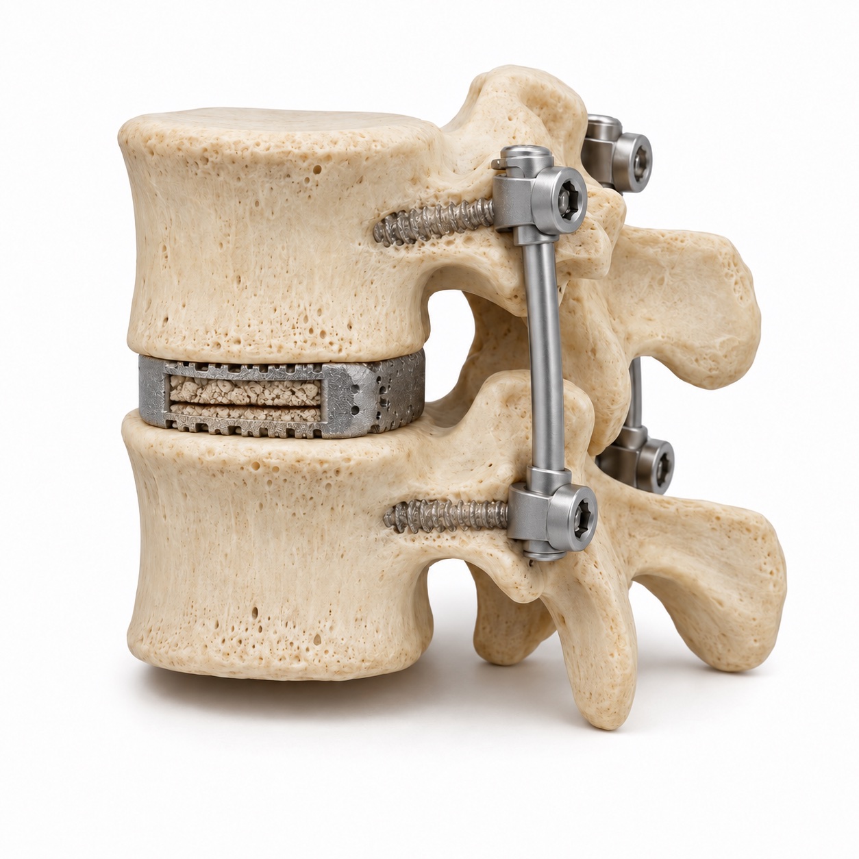
A fusion that did not form a solid bridge
Non-union concerns the level that was intended to fuse. The missing connection is represented here by a seam through the bone graft.
Hardware problems and recurrent narrowing at the original level are other possibilities. More than one problem can be present.
More about non-union, hardware problems, and recurrent stenosis
Non-union (pseudarthrosis)
A fusion asks bone to grow across a gap. When it does not, the level remains mobile, and the hardware eventually loosens or breaks under the repeated load it was never meant to carry alone. Non-union causes pain at the fused level, not the neighbor, and it usually does not produce a good interval of relief; the pain never fully settles. Dr. Boody co-authored a study of the long-term outcomes of patients without radiographic union two years after single-level ACDF, and the finding worth knowing is that some non-unions are painless and can be left alone. The ones that hurt are revised.
Hardware failure
Screws can loosen, cages can settle into soft bone, and rods can fracture. A broken rod usually means the fusion under it did not heal. Loosening around screws shows up as a halo of lucency on X-ray or CT. Hardware failure with a solid fusion is often harmless and left alone; hardware failure with a non-union is treated by addressing the non-union.
Residual or recurrent stenosis at the operated level
Sometimes the original level was not fully decompressed, or scar tissue and bone regrowth have narrowed it again. The symptoms follow the same nerve pattern as before the first surgery. The treatment is a repeat decompression at that level, which is a different operation from extending a fusion to the neighbor.
All three can coexist with true adjacent segment disease. A CT that shows both a non-union at L4-5 and a new slip at L3-4 is a plan with two parts, and that is the kind of case where experience with revision matters.
How is it diagnosed?
The history comes first: how long the first surgery helped, when the new symptoms began, and where they are. The exam maps the symptoms to a nerve root or to the cord. Comparing the new pattern with the old operative report often identifies the level before any scan.
Standing X-rays with bending views are essential. They show whether the adjacent level slips forward or opens abnormally with motion, and they show the overall alignment: whether the fusion holds a normal curve or is flat, and whether your head sits over your pelvis. CT is the test for fusion status and hardware; it shows bridging bone across the fused level, or its absence, and any lucency around screws. MRI, sometimes with metal-artifact reduction, shows the disc, the canal, and the foramina at the adjacent level, and whether the cord or root is compressed. Occasionally a CT myelogram replaces MRI when hardware obscures the picture, and a diagnostic injection at the suspected level confirms it as the pain source before a surgical decision is made.
Does alignment matter?
The lumbar spine is curved so that the body's weight passes through the discs and facets in balanced proportion. When a level is fused flat, the levels above it must hyperextend to keep the trunk upright, and that hyperextension loads their facet joints and posterior discs with every step. The same principle applies in the neck. This is why a fusion is planned with the target angle in mind, why the cage shape and the position of the screws are chosen to achieve it, and why a revision often includes correcting the alignment of the old fusion, not just decompressing the neighbor.
Alignment is also part of the motion-preservation argument. An artificial disc keeps the segment moving, but it does not automatically restore lordosis, and its long-term effect on the neighbor depends on both. Dr. Boody co-authored a ten-year comparison of adjacent-level ossification disease, the bone formation that develops next to an implant, after cervical disc arthroplasty and ACDF. Understanding what happens to the neighboring level over a decade, under both strategies, is the basis for the recommendation you get.
What are the non-surgical options?
A new disc herniation at the adjacent level behaves like any disc herniation: most improve over six to twelve weeks. Stenosis at the adjacent level often responds to an epidural injection and a therapy program aimed at flexion-based strengthening. Mechanical pain from early instability can be reduced with core strengthening and, for flares, a short course of bracing. None of these change the anatomy, but they can change the symptoms, and a level that is tolerable does not need an operation.
An injection at the suspected level has a second purpose here. Because the anatomy after a fusion is more complex and several levels may look abnormal on imaging, temporary relief from numbing one specific level confirms that it is the source before anyone plans a revision.
When is surgery considered?
The threshold for revision surgery is deliberately a little higher than for a first operation, because the operation is larger, the scar tissue raises the risk, and every extension of a fusion makes the next neighbor's job harder. Against that is the fact that nerve and cord compression do not improve with waiting, and instability tends to progress. The decision is made on your symptoms, your exam, and your imaging together, and Dr. Boody will explain where your case falls on that line.
What surgery does Dr. Boody do for it?
Read the surgical options, including hardware and alignment decisions
Extending a fusion is the most common revision. The old rods are removed, the old screws are checked and replaced if loose, new screws are placed at the adjacent level, the disc or stenosis there is decompressed, and a longer set of rods connects everything. If the old fusion is flat, the revision is the chance to correct it. In the lumbar spine, this can often be done with minimally invasive technique through the old incision or through smaller side incisions, depending on what needs to be done.
When the adjacent level is a disc herniation or foraminal stenosis without instability, a decompression alone, without extending the fusion, is sometimes enough. Dr. Boody has published on when decompression without fusion is appropriate in the lumbar spine and on far-lateral approaches that reach a herniation without disturbing the fusion. This is the least invasive option and is chosen when the anatomy allows.
In the neck, the choice between extending an ACDF and placing an artificial disc at the neighboring level is made carefully. Disc replacement next to a fusion, sometimes called a hybrid construct, keeps that level moving and is intended to reduce the stress transferred to the next level beyond it. It requires healthy facet joints, good bone, and acceptable alignment at the new level. When those are present, it is a reasonable choice; when they are not, extending the fusion is the more predictable operation. Dr. Boody's research on the long-term behavior of both is the basis for the recommendation.
What is recovery like?
Recovery is generally shorter when no new fusion needs to heal.
Early activity builds gradually while the new bone connection heals over months.
- Days 1 to 14
Begin walking and care for the incision.
Hospital stay depends on the operation. Walking starts early, with limits on bending, lifting, and twisting.
- Weeks 2 to 6
Build a comfortable routine.
Incision and muscle soreness settle. A brace or collar is used only when prescribed.
- Weeks 6 to 12
Work on strength and posture.
Therapy and lifting progression follow the surgical plan. Imaging checks the construct and early healing.
- Months 3 to 12
Let healing guide the return to activity.
Imaging helps confirm fusion. Long-term follow-up also watches the next neighboring level.
More about activity limits and recovery timing
Days 1 to 14
One to three nights in the hospital for a fusion extension; same day or one night for decompression or disc replacement. Walking from day one. Incision care and pain medication tapering. No bending, lifting, or twisting.
Weeks 2 to 6
Pain from the incision and muscle settles. Desk work resumes for most patients. Nerve symptoms from the adjacent level usually improve early; numbness lags. A brace or collar is used only when specifically prescribed.
Weeks 6 to 12
Physical therapy for core strength and posture begins. Lifting restrictions loosen. X-rays check the new hardware and early bone formation. Driving resumes once off narcotics and moving comfortably.
Months 3 to 12
Fusion is usually solid by three to six months and confirmed on X-ray or CT. Return to full activity is individualized. Long-term follow-up watches the new neighboring level with periodic X-rays.
Revision surgery carries a somewhat higher risk of complications than a first operation, including dural tears from scar tissue, infection, and non-union, and those risks are reviewed with you in detail before any decision. Your written plan will include the specific milestones for your operation.