

On this page
What is cervical myelopathy?
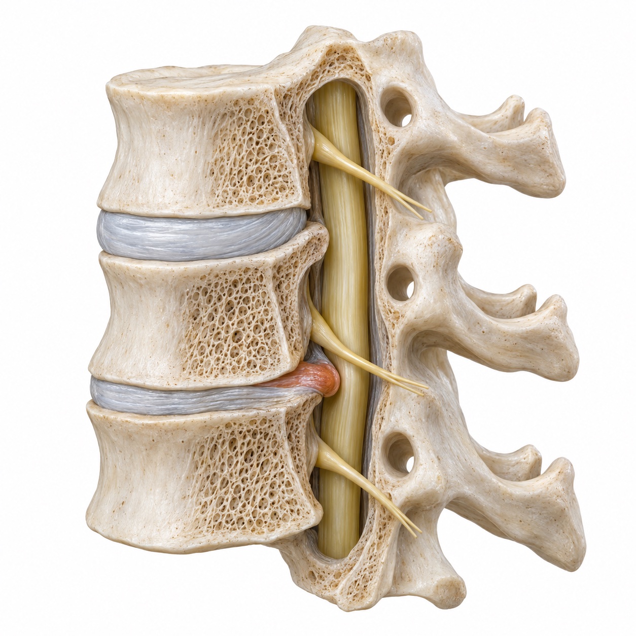
The spinal cord runs through a bony tunnel formed by the vertebrae. In the neck that tunnel has some spare room, but not much. Discs, bone spurs, and thickened ligaments can each take up part of that room, and when enough of it is gone the cord is pressed against bone with every movement of the neck. Over time the pressure and the repeated small injuries with motion disturb the cord's blood supply and its nerve fibers.
Doctors use the term degenerative cervical myelopathy when the cause is wear-related, which it usually is. It is the most common cause of spinal cord dysfunction in adults over 55. It is also frequently missed, because early symptoms are subtle and because many people expect a spine problem to hurt. Myelopathy often does not.
How is it different from a pinched nerve?
Compare the patterns
Select an area to see how the symptoms differ.
Nerve root
One arm. One path.
A pinched nerve can cause pain or numbness along a particular path in one arm.
About cervical radiculopathy- Where you notice it
- One arm, along a particular nerve’s path.
- What you may feel
- Pain or numbness along that path.
- On the exam
- Reflexes may be dampened.
Spinal cord
Hands. Legs. Balance.
Myelopathy can affect coordination on both sides, often with surprisingly little pain.
Recognize the symptoms- Where you notice it
- Both hands, the legs, and balance.
- What you may feel
- Fine-motor trouble, stiffness, or unsteadiness.
- On the exam
- Reflexes may be brisk.
Cervical radiculopathy is a wiring problem at one exit. The root is inflamed, it hurts, and once the pressure is gone it usually recovers fully. Myelopathy is a problem with the main cable. Signals traveling down to the legs and up from the hands are degraded. The classic combination is fine-motor trouble in the hands plus stiffness or unsteadiness in the legs, with brisk reflexes on exam instead of the dampened reflexes seen with a root.
The two can coexist. A disc and bone spur at C5-6 can press on the root heading to the thumb and on the cord at the same time. When that happens, the cord compression sets the plan, because it is the part that does not wait well.
What causes it?
Wear at a disc lowers its height and lets it bulge backward into the canal. The vertebral edges respond by growing bone spurs. The ligament along the back of the canal thickens and buckles inward when the neck extends. Facet joints enlarge. Each change is small, but the canal is small too, and the cord sits in the middle of all of them.
A congenitally narrow canal is common and often unknown until a scan. It leaves less reserve, so ordinary degenerative change causes compression at a younger age. A single large disc herniation can also compress the cord acutely, and a neck injury in someone with a tight canal can produce sudden myelopathy without a fracture.
OPLL deserves its own mention. The posterior longitudinal ligament runs along the back of the vertebral bodies, directly in front of the cord. In some people it calcifies and grows over years, forming a hard bar that can occupy a large share of the canal. It is more common in people of East Asian descent but occurs in every population. It changes the surgical plan because the bone is attached to the front of the canal and can be adherent to the cord's covering, and Dr. Boody has published a review of its evaluation and treatment.
What does it feel like?
- Clumsy hands: difficulty with buttons, zippers, coins, keys, or typing; handwriting that has changed
- Numbness or a glove-like tingling in the hands, sometimes on both sides
- Grip that feels weak, or dropping objects without warning
- Stiff, heavy, or unsteady legs; a wider stance; holding the rail on stairs
- Loss of balance in the dark or with the eyes closed, when you cannot use vision to compensate
- An electric sensation down the back or into the limbs when you bend the neck forward, called Lhermitte's sign
- Urgency or hesitancy with urination in later stages
- Neck pain and stiffness, which may be present but is often not the main complaint
Because the changes are slow, many people adapt without realizing it. They stop wearing shirts with small buttons, they hold the rail, they type more slowly. A useful question is whether a family member has noticed a change in the way you walk or use your hands.
Symptom check
What changes have you noticed?
Select all that apply. Your summary updates as you choose. This helps you discuss symptoms with your clinician; it is not a diagnosis.
How is it diagnosed?
The neurological exam is the heart of it. Reflexes at the knees and ankles are brisk rather than normal, and sometimes the ankle beats repeatedly when the foot is flexed, which is clonus. Flicking the tip of the middle finger makes the thumb and index finger twitch, the Hoffmann sign, a sign of an irritable cord rather than a root. The examiner may ask you to open and close your hand as fast as you can for ten seconds; fewer than about twenty cycles suggests myelopathy. Tandem gait, walking heel to toe in a straight line, is difficult. The Romberg test, standing with the feet together and the eyes closed, brings out the balance loss.
What the exam looks for
Six signs of a cord under pressure
Explore six parts of the neurological exam. Select a test to see what the clinician checks and what the finding means.
The neurological exam above describes these six signs.
How severity is measuredThe mJOA score and your baseline
Severity is often scored with the modified Japanese Orthopaedic Association scale, or mJOA, which grades hand function, walking, sensation, and bladder function out of 18. Scores of 15 to 17 are mild, 12 to 14 moderate, and 11 or below severe. The score matters because it guides the recommendation and gives a baseline to measure change against.
What MRI and other tests showImaging, signal changes, and additional tests
MRI is the key imaging test. It shows the discs, spurs, and ligament that are narrowing the canal and how much space remains around the cord. It also shows the cord itself. A bright spot within the cord on T2-weighted images, called signal change, indicates swelling or injury to the cord at that level. A dark spot on T1-weighted images suggests more established damage and a smaller chance of full recovery. CT is added to measure bone and to characterize OPLL. Flexion and extension X-rays check for instability. Nerve conduction studies and EMG are sometimes used to rule out conditions that mimic myelopathy, such as peripheral neuropathy, ALS, or multiple sclerosis. The labeled cervical MRI guide shows the cord, the fluid ring around it, and the structures that narrow the canal.
Why does it tend to progress in steps?
The pattern is not a smooth slope. Most people describe periods of stability separated by steps down, and each step tends to be permanent. That is what makes the condition different from most spine problems, where waiting costs time but not function. With myelopathy, waiting can cost function that surgery will not give back.
The practical consequence is that the decision is made on the trajectory, not on the amount of pain. Someone with mild symptoms that are stable can be followed. Someone with mild symptoms that were not present a year ago has a moving target, and that is a stronger reason to act.
What are the non-surgical options?
Observation means something specific: a repeat neurological exam and, when appropriate, a repeat MRI on a schedule, with a clear list of the changes that would trigger surgery. Patients are asked to avoid forceful neck manipulation, to be careful on ladders and ice, and to report any new hand or gait change promptly. A cervical collar is occasionally used for short periods but is not a long-term treatment.
Physical therapy can improve balance and leg strength, which reduces fall risk, and can help hand function. It treats the consequences, not the cause. Anti-inflammatory medication helps neck pain when present. Steroid injections are not a treatment for myelopathy; an injection into a narrowed canal around a compressed cord is avoided.
Current guidelines from AO Spine and the Cervical Spine Research Society recommend surgery for moderate and severe myelopathy, and either surgery or a structured program of observation and rehabilitation for mild disease, with surgery if it progresses. That matches how Dr. Boody practices.
When is surgery considered?
This is the part that surprises people. In most of spine surgery, pain drives the decision. In myelopathy, the decision is driven by function and by the risk of losing it. A patient with no neck pain, clumsy hands, brisk reflexes, and a cord that is flattened with signal change on MRI has a strong indication for surgery, because the natural course is downhill and because outcomes after decompression are better when symptoms have been present for a shorter time and are milder at the start.
What surgery reliably does is stop the progression. What it often does is improve function; on average, patients gain two to three points on the mJOA scale, and many gain more. What it does not reliably do is restore a cord that has been badly damaged for years. That is the argument for not waiting until the symptoms are severe.
Cord compression without symptoms
A specific question comes up often: what about the person whose MRI shows cord compression, even with signal change inside the cord, but who has no symptoms and a normal exam? Dr. Boody published on exactly this question in Clinical Spine Surgery in 2019. The short answer is that prophylactic surgery is not routinely recommended for asymptomatic compression, because most such patients do not develop myelopathy in the near term. The longer answer is that these patients need to be told what to watch for and followed closely, that signal change and the presence of radiculopathy at the same level raise the likelihood of developing myelopathy, and that the threshold for surgery becomes low as soon as any sign appears. This is an individualized conversation, and it is one he has often.
Where does this leave the decision?
How has it been graded?
Compared with a year ago, has it changed?
Answer both questions to see what is usually advised.
What surgery does Dr. Boody do for it?
When the compression comes from the front, from one to three discs and their spurs, an anterior approach removes it directly. ACDF takes out the disc and spurs at each level and fuses the level with a graft and plate. If the compression is behind the vertebral body itself, as with OPLL or a large spur, part of the body is removed too, which is a corpectomy, and a longer graft or cage bridges the gap. Anterior surgery also corrects a neck that has lost its normal forward curve, which matters because a cord draped over a kyphotic spine stays compressed even after posterior bone is removed.
When the compression involves three or more levels, comes mostly from the back, or the canal is congenitally narrow, a posterior approach is often better. Laminoplasty hinges the lamina open on one side and props it there with small plates, which widens the canal while preserving motion and avoiding a fusion. It requires that the neck still have a neutral or forward curve so the cord can drift backward away from the front-sided compression. Laminectomy and fusion removes the lamina outright and stabilizes the levels with screws and rods. It is chosen when there is instability, a kyphotic neck, or significant neck pain, and when preserving motion is less important than achieving a stable, decompressed canal.
Some patients need both a front and a back operation, in the same anesthetic or staged. Dr. Boody will draw the plan on your MRI and explain which approach fits your anatomy. All of these are operations he teaches to spine fellows.
What is recovery like?
Walking from the first day, often with therapy in the hospital. Anterior surgery brings a sore throat and swallowing difficulty for a week or two; posterior surgery brings more muscle soreness in the neck and shoulders. A collar may be used briefly after fusion.
Wound healed, pain medication tapered. Balance and hand coordination may already feel steadier. No lifting beyond about ten pounds. Desk work resumes for many patients in this window.
Physical and occupational therapy for gait, balance, and fine motor skills. Restrictions ease. X-rays confirm implant position and early fusion. Driving resumes once neck rotation is comfortable and narcotics are stopped.
Fusion is usually solid by three to six months. Cord recovery continues for up to a year and sometimes beyond. Most of the improvement you will get is present by twelve months. Long-term follow-up watches the adjacent levels.
How much you recover depends most on how severe the myelopathy was at surgery and how long it had been present. That is the reason Dr. Boody does not wait for severe symptoms before recommending decompression. Your recovery plan will be written down, with the milestones that apply to your operation.