

Which level is it? The dermatome map
Explore the map
Pick a level, or point to where you feel it
Choose C5 to C8 on the left, or click the part of the arm or hand where the pain, tingling, or weakness is. The figure is viewed from behind. Root symptoms are almost always one-sided, so the regions are marked on one arm.
Roots overlap and wiring varies a little from person to person. The exam, not this map, settles the level.
Compare all four nerve levelsView the map as a table: symptoms, weakness, and reflexes
| Root | Where you feel it | What gets weak | Reflex |
|---|---|---|---|
| C5 | Top of the shoulder and outer upper arm | Deltoid, lifting the arm out to the side; some biceps | Biceps |
| C6 | Thumb and index finger, outer forearm | Biceps, bending the elbow; wrist extension | Brachioradialis, biceps |
| C7 | Middle finger, back of the forearm | Triceps, straightening the elbow; wrist flexion, finger extension | Triceps |
| C8 | Small finger and inner forearm | Finger flexion and grip; small muscles of the hand | None reliable |
The map is not perfect. Roots overlap, some people have slightly different wiring, and a large herniation can catch two roots. But it is reliable enough that when the MRI and the map disagree, the map usually wins and the MRI finding is treated as incidental.
What is cervical radiculopathy?
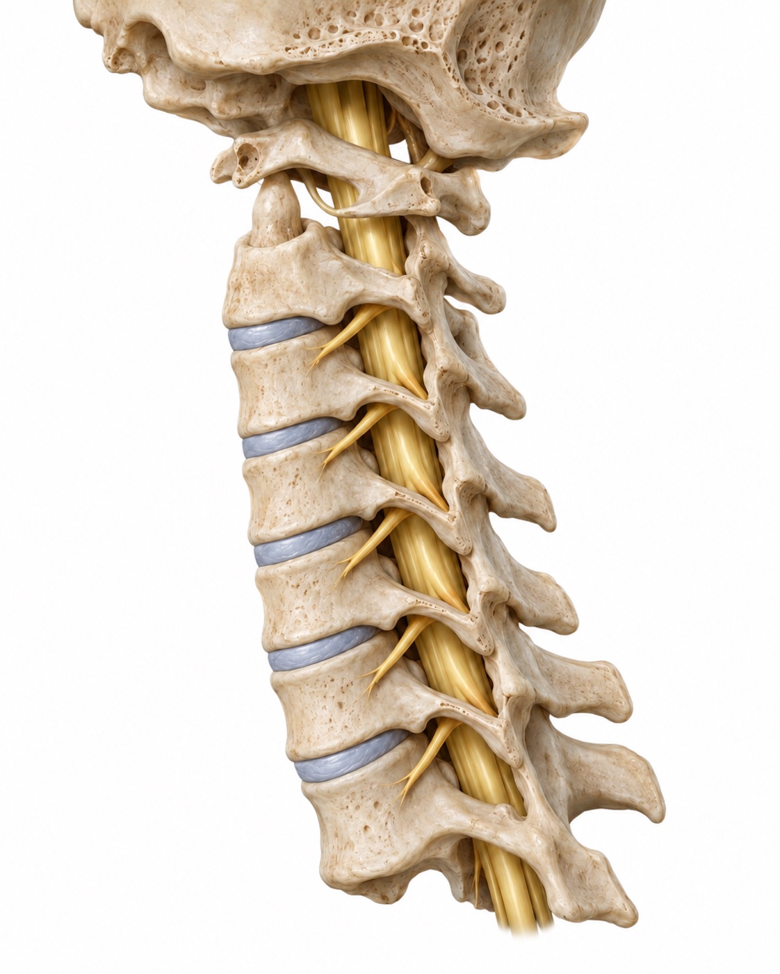
Your spinal cord runs down a bony canal in the neck. At each level, a pair of nerve roots branches off and exits through a side opening called the foramen. Those roots join together to become the nerves of the shoulder, arm, and hand. A nerve root that is squeezed or chemically irritated where it exits does not just hurt at the neck. It sends pain, pins and needles, or numbness to the patch of skin it supplies and can weaken the muscles it controls.
That mapping is the key to the whole condition. Because each root supplies a predictable strip of the arm, the location of your symptoms tells the examiner which level is involved before any scan is ordered. The most commonly affected roots are C7 and C6, which is why so many people with a pinched nerve in the neck describe pain running to the thumb, the index finger, or the middle finger.
Radiculopathy is a nerve root problem. It is different from cervical myelopathy, which is pressure on the spinal cord itself and affects coordination, balance, and both hands. If your symptoms include clumsy hands on both sides or an unsteady walk, read the cervical myelopathy page as well.
What causes it?
A cervical disc is a cushion between two vertebrae with a soft center and a tougher outer ring. When part of that center pushes through the ring toward the side, it can press directly on the exiting root. This is a soft disc herniation. It tends to come on over days, sometimes after a night of awkward sleep or a minor strain, and it is the usual cause in younger adults.
With age, the disc loses height and the small joints beside it, the uncovertebral and facet joints, form bone spurs. Together these shrink the foramen from several directions at once. This is foraminal stenosis from cervical spondylosis, and it is the usual cause after middle age. It tends to build gradually and can flare with activity or with the head held in extension.
Less common causes include a synovial cyst from a facet joint, a tumor, or an infection. These are rare, but they are part of why imaging matters when symptoms do not follow the expected course.
What does it feel like?
A typical checklist looks like this. You do not need every item, but the pattern of a one-sided arm symptom that follows a path is what matters.
- Pain that runs from the neck or between the shoulder blades into the shoulder, arm, or hand, almost always on one side
- Tingling, pins and needles, or numbness in a specific part of the hand, such as the thumb, the middle finger, or the small finger
- Weakness in a specific movement: lifting the arm, bending the elbow, straightening the elbow, or gripping
- Pain that is worse when you look up, turn toward the painful side, or hold the head still for a long time, and sometimes better with the hand rested on top of the head
- Pain that wakes you at night or makes it hard to find a comfortable head position
- Neck stiffness or muscle spasm, which often accompanies the arm pain but is not the main problem
Weakness deserves special attention. Pain and tingling are unpleasant, but they usually recover completely. Weakness that is measurable on exam, or a reflex that has gone missing, means the root is under enough pressure to affect its motor fibers, and that shortens the time you should spend waiting.
Check the pattern
Does this sound like a pinched nerve?
Tick what applies. You will get a plain reading of the pattern and what usually comes next. It is reading, not a diagnosis.
Tick any items above to read the pattern.
How is it diagnosed?
The exam checks strength in each muscle group, sensation in each dermatome, and the reflexes at the biceps, brachioradialis, and triceps. A few provocative maneuvers help. In the Spurling test, the head is tilted toward the painful side and gently pressed down; reproduction of the arm pain points to the foramen. Relief when you rest your hand on your head, the shoulder abduction sign, points the same direction. The exam also screens for signs of spinal cord involvement, because that would change the urgency.
Plain X-rays show disc height, bone spurs, alignment, and, on flexion and extension views, whether one vertebra slips on the next. They do not show nerves. MRI is the test that shows the soft tissues: a disc herniation, the size of the foramen, and whether the spinal cord is also being touched. On the MRI report you may see the words "foraminal narrowing," "disc protrusion," or "uncovertebral hypertrophy" at the level that matches your symptoms. To see what those structures look like, open the labeled cervical MRI guide.
A CT scan shows bone in more detail and is useful when surgery is planned or when an MRI cannot be done. A CT myelogram, in which contrast is injected into the spinal fluid before the scan, outlines the nerves when MRI is not possible. Nerve conduction studies and EMG are added when the story is not clear, most often to separate a cervical root from carpal tunnel syndrome, ulnar nerve entrapment at the elbow, or a shoulder problem, all of which can mimic radiculopathy.
What are the non-surgical options?
The natural history is favorable. Published series report that a large majority of people with cervical radiculopathy improve substantially without an operation, most within the first three months. That is the reason surgery is not the first conversation for someone whose main symptom is pain.
In the first two weeks, the goal is comfort. A soft collar for a few days can help sleep but should not be worn for long. Non-steroidal anti-inflammatory medication, when your stomach and kidneys allow it, reduces the swelling around the root. A short oral steroid taper is sometimes used for severe pain. Positions that open the foramen, such as slight forward flexion and a hand on the head, are used freely.
After the acute phase, physical therapy addresses posture, gentle cervical traction, nerve gliding, and strengthening of the muscles that stabilize the neck and shoulder blades. Therapy does not shrink a disc herniation, but it reduces the mechanical load on the irritated level and shortens the time to recovery for many patients.
If pain persists beyond a few weeks, a cervical epidural steroid injection or a selective nerve root injection delivers anti-inflammatory medication directly to the root under X-ray guidance. Roughly half of patients get meaningful relief, which can be enough to allow the herniation to resolve on its own. The injection also serves as a diagnostic test: if numbing the suspected root eliminates your pain, the level is confirmed.
When is surgery considered?
Three situations move surgery up the list. The first is failure of time and treatment: you have done the therapy and the injection, three months have passed, and the arm pain still limits work or sleep. The second is progressive motor weakness, for example a triceps that is losing strength on serial exams. Nerves recover better when the pressure is removed before the damage becomes fixed. The third is the presence of cord compression or early myelopathy on the same scan, which changes the question from comfort to protection.
What surgery is not for is neck pain alone. Decompressing a root treats root symptoms. Someone with axial neck pain and an MRI that shows a bulging disc, but no arm symptoms and a normal exam, is not helped by root decompression and is generally not offered it.
Where are you in the course?
How long have the arm symptoms been going on?
Is there weakness?
Answer both questions to see where you are.
What surgery does Dr. Boody do for it?
In an ACDF, the disc is removed through a small incision at the front of the neck, the root and cord are decompressed under the microscope, and a spacer with bone graft is placed where the disc was. A small plate holds it while the two vertebrae fuse into one. ACDF has been the standard operation for more than sixty years, and its results for arm pain are consistent and durable. Its trade-off is that the fused level no longer moves, which puts slightly more work on the levels above and below.
Cervical disc replacement uses the same approach and the same decompression, but places an artificial disc that preserves motion at the level. Randomized trials with long follow-up have shown that it relieves arm symptoms as well as fusion, with lower rates of reoperation at adjacent levels in most series. It is a good option when the facet joints at the level are healthy, the bone quality is good, and the alignment is normal. It is not the right choice for every patient, and Dr. Boody will tell you which category you fall into and why.
A third option, posterior cervical foraminotomy, opens the foramen from the back of the neck through a small incision without removing the disc or fusing anything. It suits a herniation that sits far to the side. It does not address bone spurs in front of the root and is used selectively.
All three are typically single-night or same-day procedures. Most patients notice that the arm pain is gone or much better when they wake up, because the pressure that caused it has been removed.
After surgery
What is recovery like?
What improves first
Arm pain usually improves first, often within days. Numbness and weakness recover more slowly, over weeks to months, because the nerve has to heal.
Healing after surgery
After ACDF, the fusion itself takes three to six months to become solid; after disc replacement there is no fusion to wait for, but the soft tissues still need about six weeks.
Days 1 to 14
Right after surgery
- Home the same day or the next morning.
- A sore throat and some difficulty swallowing are common for a week or two.
- Walking is encouraged from the first day.
- No lifting over about ten pounds.
- Most people do not need a collar.
Weeks 2 to 6
Early healing
- Desk work often resumes within one to two weeks; driving once you are off narcotic pain medication and can turn your head comfortably.
- Neck stiffness eases.
- Numbness may still be present; that is expected.
Weeks 6 to 12
Building strength
- Physical therapy for posture and strength if needed.
- Lifting restrictions loosen.
- Follow-up X-rays check the position of the implant.
- Most physical jobs restart during this window, with modifications for fusion patients.
Months 3 to 12
Full recovery
- A fusion is usually solid by three to six months and confirmed on X-ray.
- Nerve recovery continues for up to a year, so residual numbness at three months can still improve.
- Return to unrestricted activity, including contact sports, is individualized.
Your recovery plan
Recovery ranges depend on the number of levels, whether it was a fusion or a replacement, your general health, and how long the nerve was compressed before surgery. Dr. Boody will give you a timeline that fits your operation, and it will be written down in your plan.