

What is spine trauma?
The spine is a stack of vertebrae connected by discs in front and by paired facet joints and strong ligaments behind. Together they protect the spinal cord and let you move. A fracture breaks one or more of the bones; a ligamentous injury tears the soft tissue that holds the bones in line. Some injuries do both. The consequence that matters is not the crack itself but whether the spine can still hold its shape and protect the nerves under normal loads.
Spine trauma spans a wide range. At one end is a mild compression fracture in an older adult that heals in a few weeks with pain control. At the other is a fracture-dislocation with spinal cord injury that requires urgent surgery. Most injuries sit somewhere between, and sorting out where is the job of the first days after the injury.
Is the spine stable?
Can the injured bones and ligaments hold their alignment under load? This helps determine whether the injury can heal with non-surgical care or needs stabilization.
Are the nerves affected?
The examination and scans look for pressure on the cord or nerves. Weakness, numbness, or bowel or bladder changes make the evaluation urgent.
See the difference
What changes when a vertebra breaks?
Compare two fracture patterns with healthy bone. Look at the middle vertebra, especially its front edge and the back wall beside the spinal canal.
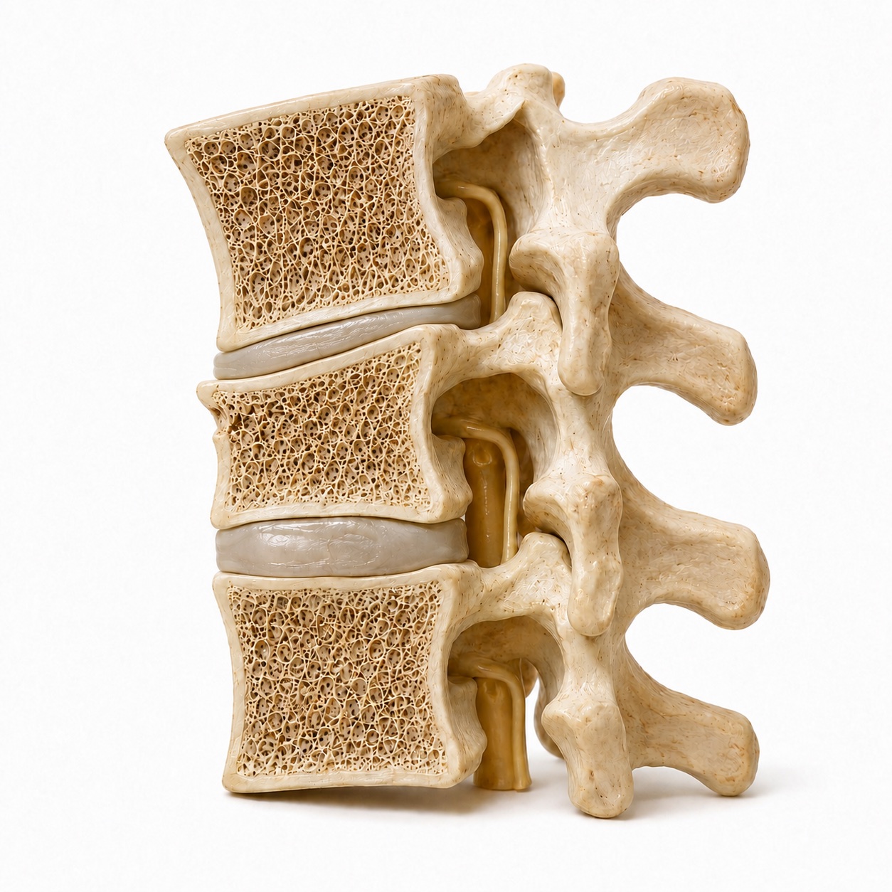
Compression fracture
The front collapses into a wedge.
The front of the vertebral body loses height while the back remains intact. This can happen after a minor fall when osteoporosis has weakened the bone.
- What it can mean for treatment
- Usually stable. Many heal with pain control, activity adjustment, and sometimes a brace. A low-energy fracture also calls for a bone-health evaluation.
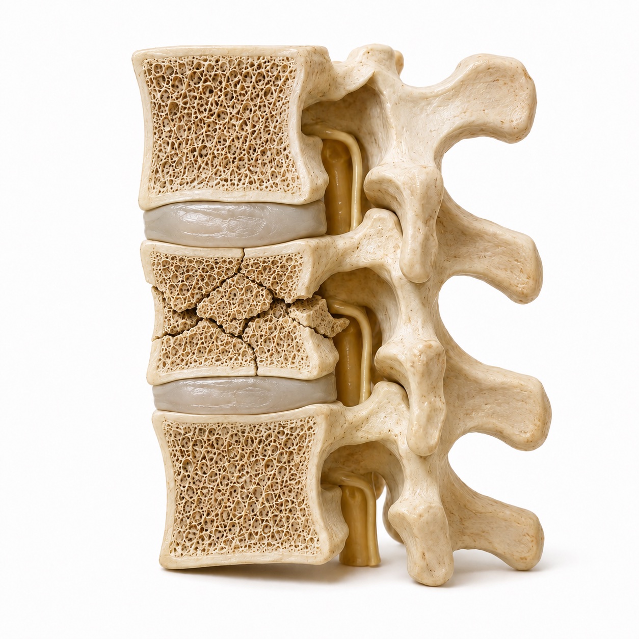
Burst fracture
The fracture reaches the back wall.
Both the front and back of the vertebral body break. Fragments can spread outward and push backward toward the cord or nerves.
- What it can mean for treatment
- Some are stable; others need surgery. The ligaments, alignment, and neurological exam help Dr. Boody decide whether observation or stabilization is appropriate.
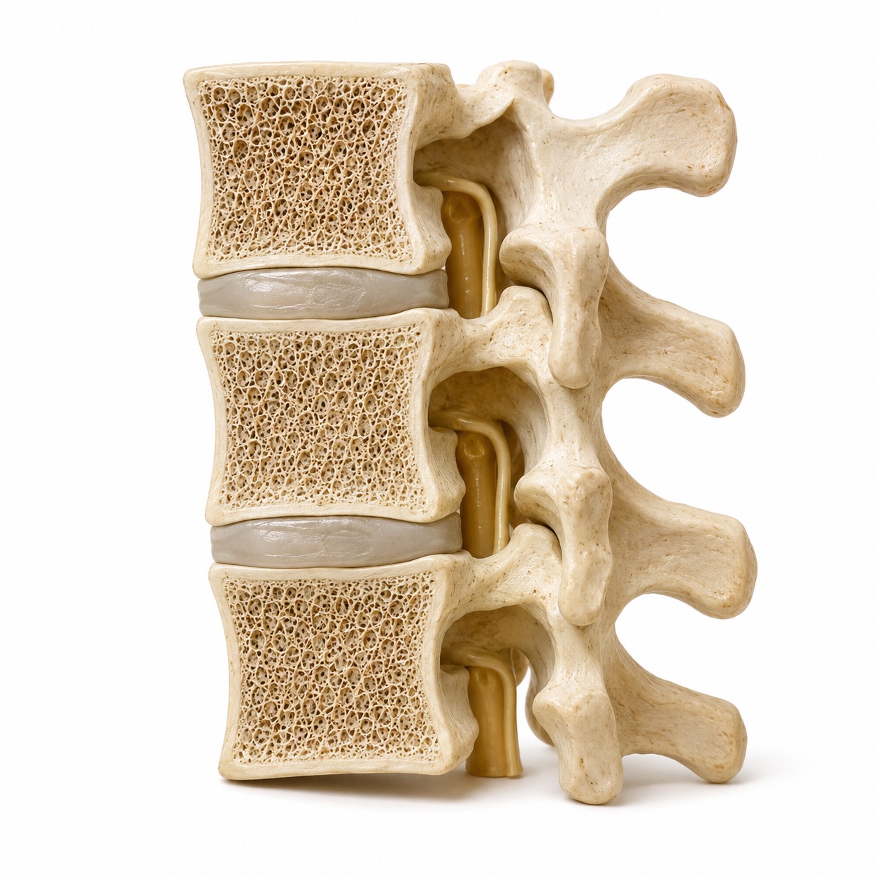
Healthy comparison
The bone keeps its height and shape.
Compare the middle vertebra with the two fracture examples. Its front and back walls are intact, and the discs separate it from the bones above and below.
- Why bone health matters
- After a fracture from a minor fall, care includes looking for osteoporosis and treating weakened bone to help prevent another fracture.
Educational illustrations of the mid-back and low-back spine. Your examination and imaging determine the fracture pattern and stability.
What causes it?
A straight downward load, as in landing on the feet or the buttocks, crushes the front of a vertebral body and produces a compression or burst fracture. A forward-bending force with the body restrained, as in a lap belt in a crash, can tear the spine open from the back, a flexion-distraction injury. A twisting or shearing force can dislocate one vertebra on another, the most unstable pattern. In the neck, a blow to the top of the head can drive the skull down onto the upper cervical spine, and hyperextension can fracture the odontoid, the peg of bone that the skull pivots on.
Osteoporosis changes the equation. Bone that has lost density can fracture from a sneeze, lifting a grandchild, or a fall from standing height. These fractures are common, they are often stable, and they are a signal to treat the bone as well as the spine, because one osteoporotic fracture predicts another.
What does it feel like?
- Sharp pain in the neck or back at one spot, worse with sitting up, standing, or turning
- Tenderness over the spine when it is pressed, sometimes with a visible step or bump
- Muscle spasm and an unwillingness to move the head or trunk
- Pain that wraps around the chest or abdomen along a rib, from an irritated nerve at a mid-back fracture
- Numbness, tingling, or weakness in the arms (neck injury) or legs (mid-back or low-back injury)
- Difficulty walking, or legs that feel heavy or do not respond normally
- Loss of bladder or bowel control, inability to urinate, or numbness in the groin and inner thighs
- In an older adult, new back pain and a loss of height or a forward stoop after a minor fall
Not every fracture hurts badly. Some osteoporotic compression fractures produce only a dull ache and are found weeks later on an X-ray taken for another reason. And a person with several injuries from a crash may not feel a spine fracture at all until the other injuries are treated, which is why trauma teams image the whole spine when the mechanism is severe.
Is the spine stable or unstable?
Stable injury
- Bone is broken but the posterior ligaments and facet joints are intact
- Vertebral body has lost less than about half its height, with little forward angulation
- No fragments pushed into the canal, or only small ones without nerve symptoms
- Normal neurological exam
- Alignment does not change between lying and standing X-rays
- Typical plan: pain control, early walking, a brace for comfort in some cases, follow-up X-rays
Unstable injury
- Posterior ligaments torn, facets fractured or dislocated, or spinous processes spread apart
- Vertebral body crushed with significant angulation or more than about half its height lost
- Bone driven into the canal with narrowing that threatens or compresses the cord
- Weakness, numbness, or bladder change on exam
- One vertebra shifted on another, or alignment that worsens when upright
- Typical plan: surgical stabilization, with decompression if the nerves are compressed
Spine surgeons use scoring systems to make this judgment consistent. For the thoracic and lumbar spine, the Thoracolumbar Injury Classification and Severity score, or TLICS, adds points for the shape of the fracture, whether the posterior ligaments are intact, and whether there is a neurological deficit. Low scores are treated without surgery, high scores with it, and the middle is a judgment call that depends on you. The AO Spine classification does similar work with a more detailed description of the fracture pattern. Both exist so that the recommendation you receive is based on the injury, not on the habits of whoever happened to see you first.
The three columns, in plain words
The front column is the front half of the vertebral body and disc, with the ligament that runs down the front. It bears most of your weight. The middle column is the back half of the body and disc, with the ligament that runs along the back of the bodies, just in front of the spinal cord. The back column is everything behind the canal: the facet joints, the lamina, the spinous process you can feel through the skin, and the strong ligaments that connect them.
A simple compression fracture crushes only the front column; the middle and back columns hold, so it is stable. A burst fracture involves the front and middle columns, which is why it can push bone backward into the canal and why it needs a closer look. A fracture-dislocation disrupts all three, and the spine is unstable by definition. When your surgeon points at the CT and talks about the posterior ligamentous complex, this is the back column, and whether it is intact is often the deciding fact.
What is a burst fracture?
The junction between the rigid thoracic spine, which is splinted by the rib cage, and the mobile lumbar spine, roughly T11 to L2, absorbs the force of a fall or crash. When a vertebral body there is loaded beyond its strength, it fails like a can crushed from the top: the walls bow out, and fragments from the back wall are pushed toward the cord and nerves. The CT shows how much of the canal is occupied and whether the back column held.
A burst fracture with an intact posterior ligamentous complex, no neurological deficit, and modest angulation is stable enough to heal without surgery in most patients, and randomized studies have found similar outcomes whether or not a brace is used. Dr. Boody co-authored a review of thoracolumbar burst fractures in Clinical Spine Surgery in 2018 that lays out this decision. A burst fracture with a torn posterior ligamentous complex, significant kyphosis, or a neurological deficit is treated surgically. Between the two is a group where the decision depends on your age, your bone, your other injuries, and how you are doing when you stand up.
How is it diagnosed?
Emergency departments now image the whole spine with CT after high-energy trauma, because a second fracture at another level is found in a meaningful share of patients. The CT shows the fracture pattern, the height loss, the angulation, and how much bone is in the canal. It also shows facet joints that are perched or locked, and spinous processes that have spread apart, which are clues that the back column has failed.
MRI shows what CT cannot: the ligaments, the discs, the cord, and the nerves. A bright signal in the ligaments behind the spine means they are torn. A bright signal in the cord means it is bruised or swollen. MRI is ordered when the neurological exam is abnormal, when the CT leaves the stability question open, or when surgery is planned and the surgeon needs to see the soft tissues.
Standing X-rays are the test many people are surprised by. A fracture that looks acceptable on a CT taken while lying flat may angulate when you stand. Dr. Boody uses upright films, in a brace when appropriate, to answer the question the CT cannot: what does this spine do when it has to carry you?
Bracing and non-surgical care
A brace does not heal a fracture; the bone does that. What a brace does is limit painful motion and remind you to move carefully during the weeks the bone is knitting. For stable thoracolumbar burst and compression fractures, studies have shown that outcomes with and without a brace are similar, so the brace is a tool for comfort and confidence rather than a requirement. When one is used, it is typically a rigid or semi-rigid thoracolumbar orthosis worn when upright for six to twelve weeks. In the neck, a hard collar serves the same purpose for stable cervical fractures.
Early walking matters more than most people expect. Lying in bed weakens muscle, raises the risk of blood clots and pneumonia, and slows recovery. The goal in the first days is to be up and moving with the brace on and pain managed. Physical therapy teaches log-rolling out of bed, safe bending, and gradually progressive activity.
For osteoporotic compression fractures that remain very painful after a few weeks, vertebral augmentation, injecting bone cement into the fractured body, can reduce pain and is considered case by case. Every low-energy fracture also prompts a bone density test and a conversation about treating osteoporosis, because the next fracture is the one you can still prevent.
When is surgery considered?
The purpose of surgery in trauma is different from most elective spine surgery. It is not primarily to relieve pain. It is to restore the spine's ability to protect the nerves and carry load so that healing can happen in the right position. That means realigning the fracture, holding it with screws and rods, and, when needed, removing bone from the canal to free the cord or nerves.
Timing depends on the situation. A fracture-dislocation with spinal cord injury is operated on urgently. An unstable burst fracture without a deficit is scheduled within days, once other injuries are addressed and the patient is medically ready. A fracture that was stable at first but is angulating on follow-up X-rays is operated on when that change is recognized, which is why the follow-up films are not optional.
What surgery does Dr. Boody do for it?
Percutaneous pedicle screw fixation places screws into the vertebrae above and below the fracture through incisions about the width of a finger, guided by X-ray or navigation, and connects them with rods slid under the skin. The muscles are spread rather than cut. For a burst fracture with intact ligaments in a patient who needs stabilization, this acts as an internal brace: it holds the alignment while the bone heals and can be removed after a year if desired, with no fusion. Blood loss is small, hospital stays are short, and patients are up the next day. Dr. Boody has published on the training of pedicle screw placement and teaches the technique to fellows.
When the posterior ligaments are torn or the fracture is a dislocation, the levels are fused as well, because ligaments do not heal reliably enough to trust the spine's stability once the hardware is gone. When bone is compressing the cord or nerves, it is removed, from the back through the pedicle or from the front through the chest or abdomen, and the gap is rebuilt with a cage.
In the neck, the pattern decides the approach. An odontoid fracture may be fixed with a single screw from the front or fused to the vertebra below from behind. A facet dislocation is reduced, sometimes with traction, and fused. A cervical burst fracture is often treated with removal of the vertebral body from the front and a cage and plate, sometimes with a posterior fusion added. Dr. Boody performs all of these and will show you, on your CT, why one is chosen over another. See the full list of treatments.
Spinal cord injury: what timing means
The cord is injured twice: once by the mechanical force at the moment of impact, and again over the following hours by swelling, bleeding, and loss of blood flow in a canal that is now too small. Surgery cannot undo the first injury. It can limit the second by removing pressure and restoring stability so the cord can be perfused and rested. That is the reason for urgency.
The neurological exam is graded on the American Spinal Injury Association scale from A, complete, with no motor or sensory function below the level, to E, normal. An incomplete injury, in which some sensation or movement is preserved below the level, has a much better chance of meaningful recovery, and the exam is repeated often in the first days because it changes. High-dose steroids, once routine, are no longer given routinely; they are discussed in specific situations within the first hours of injury. Blood pressure is kept in a target range in the intensive care unit to protect cord blood flow. Rehabilitation begins as soon as the spine is stable.
If you were seen in an ER: what to bring
- Your imaging. Ask the emergency department or the hospital's radiology department for a disc or a portal link with the CT scan and any MRI or X-rays. Reports alone are not enough; Dr. Boody needs to see the images.
- Your discharge papers. They list the diagnosis, the medications you were given, and the restrictions you were told to follow.
- Your brace. Wear it as instructed and bring it even if you have stopped using it, so its fit and type can be checked.
- A list of what has changed. Any new numbness, weakness, bladder or bowel change, or a pain that has moved or worsened since the ER visit.
- Your medication list and other diagnoses, especially blood thinners, osteoporosis, and prior spine surgery.
- Someone to drive. You may have standing X-rays taken and should not be driving in a rigid brace.
If your symptoms worsen before the appointment, go back to the emergency department rather than waiting. Call the office at (317) 228-7000 to be seen sooner; new fractures are fit into the schedule.
What is recovery like?
Up and walking with the brace or after surgery, with pain managed and therapy teaching safe movement. Hospital stays after percutaneous fixation are typically one to three nights. Standing X-rays confirm alignment before discharge.
Brace worn when upright if prescribed. No bending, lifting, or twisting. Walking daily, increasing distance. Follow-up X-rays at about two and six weeks watch for any change in alignment. Desk work often resumes.
X-rays or CT show early healing. The brace is weaned. Physical therapy begins strengthening of the core and back. Driving resumes when the brace is off and pain medication is stopped.
Bone is solid by three to six months. Restrictions lift progressively. Percutaneous screws placed without fusion may be removed after about a year. Neurological recovery continues throughout this period.
Ranges are wide because injuries are. A single compression fracture in a healthy adult sits at the fast end; a multilevel injury with cord involvement sits at the slow end and includes inpatient rehabilitation. You will have a written plan that fits your injury, with the follow-up dates and the reasons for each.