

What is minimally invasive spine surgery?
See the difference
Two routes to the same target.
Compare how the surgeon reaches the spine. The best route depends on the view and access your operation needs.
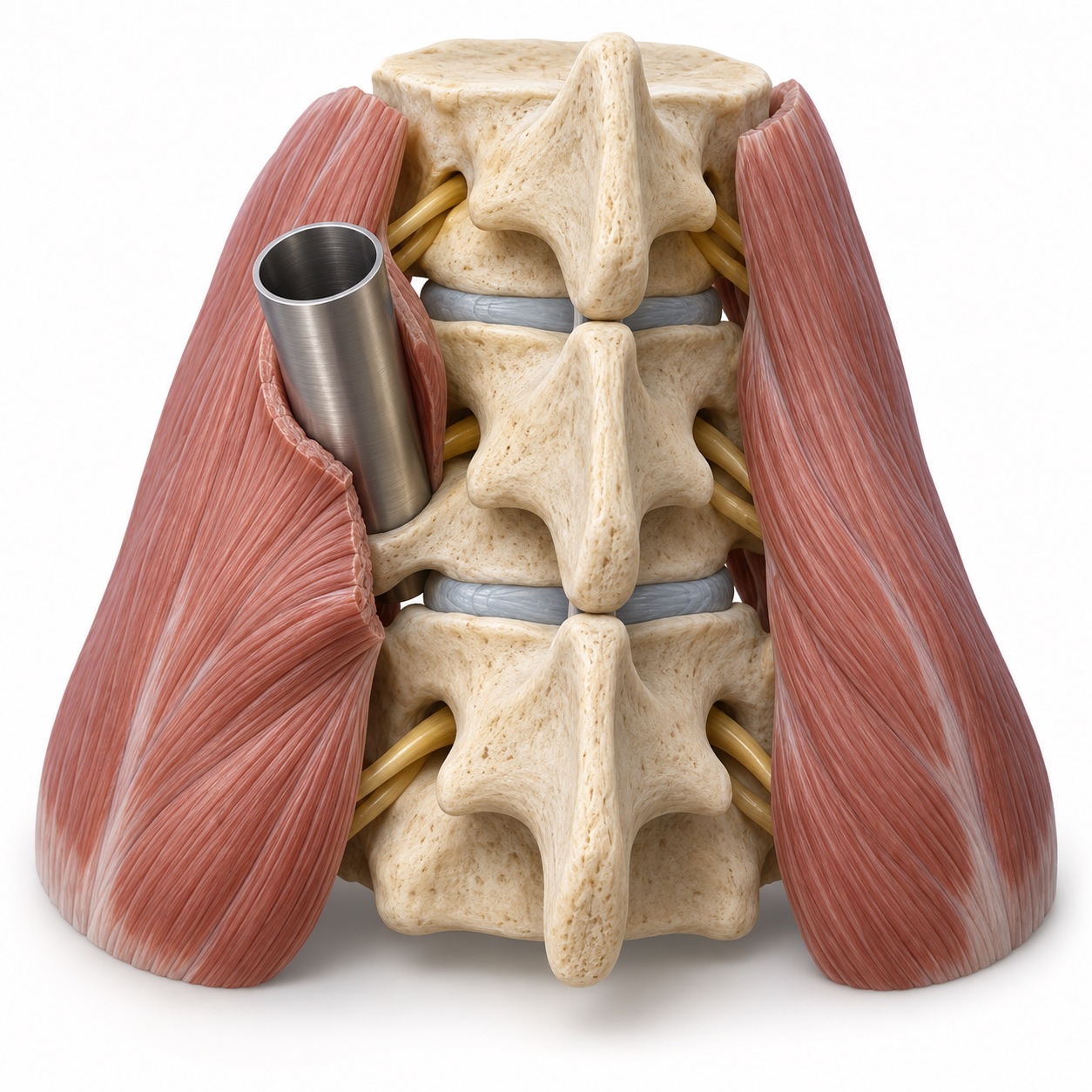
Through a narrow tube
Muscle fibers are spread to create a focused working corridor. The surgeon uses magnification to work through the tube.
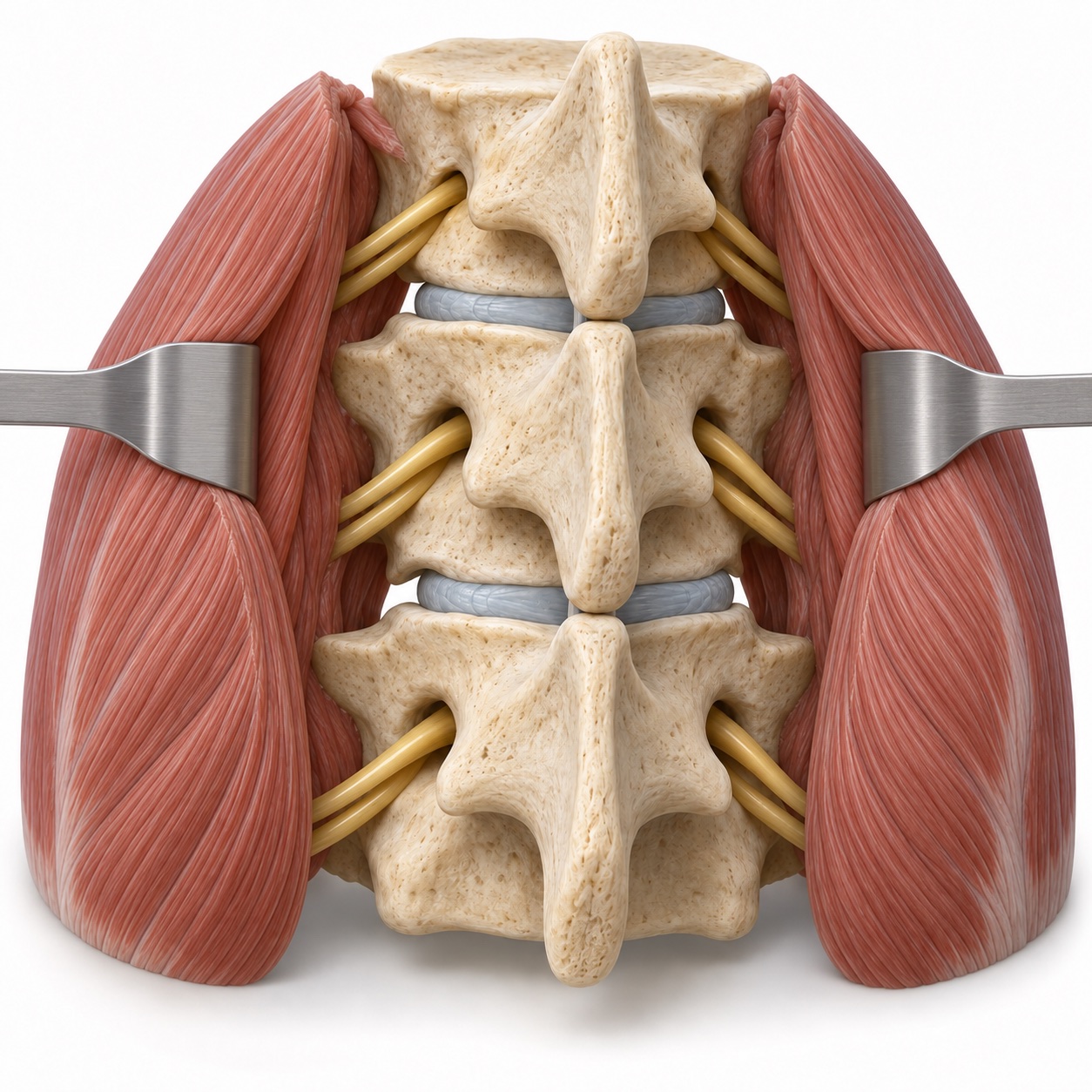
Through a wider opening
Muscle is moved away from the back of the spine to expose a broader area. This can provide the view needed for more extensive work.
Illustrative anatomical models; tissue and access are simplified. A smaller opening does not change the need to treat the whole problem safely.
Read more about the technique and the five procedures Dr. Boody performs
In a traditional open lumbar operation, the incision runs along the midline and the muscles on either side are detached from the spine and held back for the whole procedure. That gives a wide view, but the muscle is the part of the operation that hurts most afterward and heals slowest.
In a minimally invasive approach, a small incision, usually about an inch, is made off to the side of the midline. A series of dilators is passed through the muscle to spread the fibers apart rather than cut them, and a tube or an expandable retractor is placed over them. The surgeon works down the tube under a microscope or with magnifying loupes, with X-ray or navigation confirming the position. The muscle closes back over the track when the tube is removed.
Dr. Boody uses this approach for five lumbar problems in particular:
- Far-lateral (extraforaminal) microdiscectomy, for a disc herniation that sits outside the nerve tunnel and pinches the nerve that has already exited. Dr. Boody authored the technique paper on this approach in Clinical Spine Surgery (2021).
- Decompression of lumbar facet cysts, fluid-filled sacs that grow from a worn facet joint into the canal and pinch a nerve. His 2021 paper asked whether cysts without instability need fusion at the time of decompression.
- Tubular decompression for spinal stenosis, opening a narrowed canal from one side while preserving the midline bone and ligaments.
- Minimally invasive TLIF, a fusion between two vertebrae done through the same small corridor when the level is unstable, most often for spondylolisthesis. He led a prospective study of TLIF with a bone graft extender, published in 2019.
- Percutaneous pedicle screw fixation, screws and rods placed through small stab incisions to stabilize the spine, including after fractures, without opening the back.
Who is minimally invasive surgery for, and who is it not for?
The decision is about the problem, not about the size of the incision. A far-lateral herniation is a classic minimally invasive target because the nerve is pinched in a spot that an open midline approach reaches poorly anyway. A single-level facet cyst or a one-level stenosis is another. A one-level slip that needs a fusion can usually be done as a minimally invasive TLIF. A stable-enough burst fracture that needs a brace-like internal support can often be stabilized with percutaneous screws.
Multi-level stenosis, scoliosis or other deformity needing correction, tumors, infections needing wide debridement, and many revision cases with dense scar are better served by an open approach that gives a full view. Body habitus can go either way: in larger patients the narrow tube can actually reduce the depth of the wound, but very deep tissue may also limit what can be seen. Dr. Boody will tell you which category you are in and why.
Whichever approach is used, surgery is offered only after the same threshold has been met: symptoms that have not improved with time and non-surgical care, or weakness or nerve compression that should not wait.
What happens during the operation?
Inside the spine
What is the operation trying to do?
Relieving nerve pressure and stabilizing a level are different goals. Select a view to see what each involves.
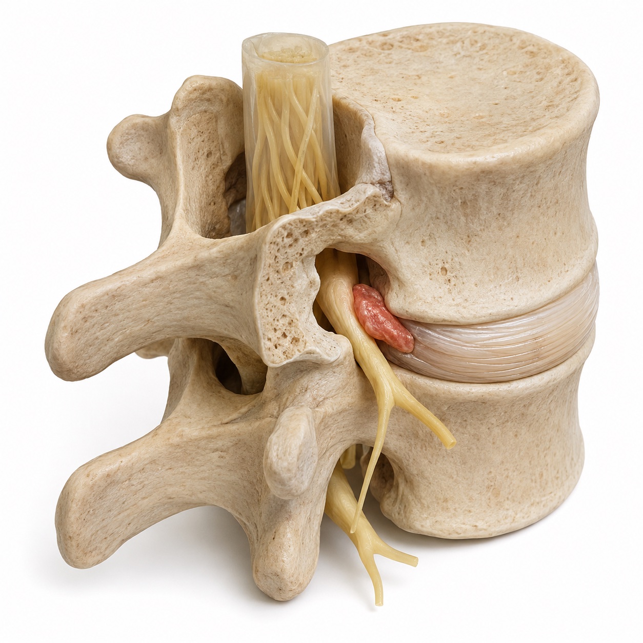
Make room for the nerve
The red area illustrates disc material crowding a nerve. Decompression removes the tissue causing pressure. A disc fragment, a cyst, or excess bone can be the target.
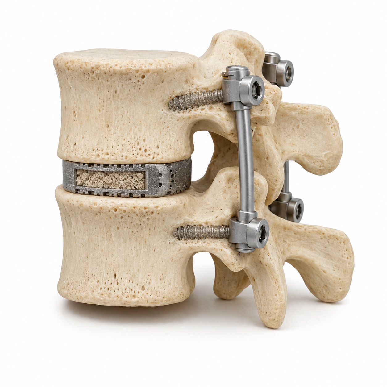
Support a level while it fuses
When fusion is needed, a cage with bone graft sits between the vertebrae. Screws and rods support the level while the bone heals.
These models explain the treatment goals, not a particular patient’s operation. The disc image shows one example of nerve pressure; fusion hardware and placement vary.
Walk through the surgical steps, including fracture fixation
- Anesthesia and positioning. You are fully asleep and positioned face down on a padded frame that lets the abdomen hang free, which reduces bleeding. X-ray is used to mark the level on the skin before the incision.
- Incision and corridor. A one-inch incision about two finger-widths from the midline. Dilators of increasing size are passed to the bone, then a tube or expandable retractor is docked over them. For a far-lateral herniation the corridor is angled further out, between two muscle groups, to reach the nerve outside the tunnel without removing the facet joint.
- Decompression. Under a microscope, a small window of bone and ligament is removed to expose the nerve. For a herniation, the loose fragment is lifted out and the disc is cleaned of any other loose pieces. For a cyst, the cyst is separated from the nerve covering and removed with the ligament it grew from. For stenosis, the surgeon works across the midline from one side to open both sides of the canal.
- Fusion, when needed (TLIF). The facet joint on one side is removed to reach the disc. The disc is cleared and a cage packed with bone graft is placed in the space. Screws are inserted into the vertebrae above and below through the same or small adjacent incisions and connected by rods to hold the level still while it heals.
- Percutaneous screws for fractures. Through four to six small stab incisions, screws are guided into the vertebrae above and below the fracture under X-ray, and rods are slid under the skin to connect them. The fracture itself is not opened. In some patients the hardware is removed once the fracture has healed.
Operating time is one to two hours for a microdiscectomy, cyst removal, or one-level decompression, and two to four hours for a one-level TLIF or a fracture fixation. Microdiscectomy and decompression patients usually go home the same day or after one night. TLIF patients typically stay one to two nights. Fracture patients stay one to three nights depending on the injury.
What are the alternatives?
Injections. An epidural steroid injection can calm an inflamed nerve. A facet cyst can sometimes be drained or ruptured under imaging guidance, though recurrence is common and many patients go on to surgery.
Open surgery. The same decompression or fusion through a midline incision with the muscles reflected. It is the right choice when the field needs to be wide.
Decompression alone versus fusion. This is the decision that matters most for facet cysts and for slips. A cyst that grew from a stable joint can usually be removed without a fusion. A cyst at a level that is already slipping, or a slip that moves on bending X-rays, usually needs the level stabilized as well. Dr. Boody's 2021 paper addressed exactly that question for cysts without instability.
Compare the benefits and tradeoffs of each approach
Open surgery
- Midline incision; muscles detached from the bone and retracted
- Wide view; suits multi-level, deformity, tumor, and many revisions
- More blood loss and muscle soreness; longer hospital stay on average
- Same decompression, same fusion, same long-term goal
Minimally invasive
- One-inch incision off the midline; muscle split, not stripped
- Narrow view under magnification; suits well-defined one- or two-level problems
- Studies report less blood loss, less early pain, shorter stays
- More X-ray use during surgery; not appropriate for every problem
What are the risks?
- Dural tear. The thin membrane around the nerves can be nicked, especially when there is scar from prior surgery or when a cyst is stuck to it. Studies report this in a small percentage of first-time cases and more often in revisions. It is repaired or sealed during the operation and usually has no lasting effect, but may mean a day or two of lying flat.
- Recurrent herniation. After any microdiscectomy, a new fragment can herniate from the same disc. Studies report recurrence in roughly 5 to 15 percent of patients over the years after surgery, more often with a large tear in the disc wall.
- Incomplete decompression. The narrow view raises the chance that a fragment or a ridge of bone is missed. Careful preoperative planning and intraoperative X-ray lower it.
- Nerve injury. Uncommon. Temporary numbness or weakness is more likely than permanent injury. Far-lateral surgery works close to the exiting nerve and its small ganglion, which can be irritated for weeks.
- Screw position and non-union. For TLIF and percutaneous fixation, a screw can be placed close to a nerve or outside the bone and need repositioning. A fusion can fail to heal, more often in smokers. Studies report fusion rates for single-level TLIF in the high range, but not one hundred percent.
- Infection, blood clots, anesthesia. All low. Several studies report lower infection rates with minimally invasive approaches, likely because less tissue is exposed for less time.
- Radiation. Minimally invasive surgery relies on X-ray during the operation. The dose to you is modest and is tracked.
- Conversion to open. Occasionally the view through the tube is not safe enough, and the operation is converted to an open approach. This is a judgment call made for your safety, not a complication.
What is recovery like?
Often home within a day, with a return to desk work in one to two weeks.
Bone healing takes months, with lifting restrictions for longer.
-
First week
Start with short walks.
Walking usually begins the day of surgery. Expect incision soreness and limits on bending, lifting, and twisting.
-
Weeks 2 to 6
Build your daily routine.
Walking distance increases. Desk work often resumes earlier after decompression than after fusion.
-
Months 2 to 3
Rebuild strength and movement.
Physical therapy focuses on core strength and body mechanics. Fusion patients progress with lifting limits still in place.
-
Months 3 to 12
Return as healing allows.
After fusion, heavier lifting and impact wait until healing is confirmed. Follow-up imaging helps guide the next step.
More about activity limits and recovery timing
First week
Walking from the day of surgery, several short walks a day. Incision soreness rather than deep muscle pain. No bending, lifting over about ten pounds, or twisting. Leg pain from a pinched nerve often improves immediately; numbness lags.
Weeks 2 to 6
Desk work by one to two weeks for decompression, two to four weeks for fusion. Driving once off narcotic medication. Walking distance builds up. After a fusion, a brace is sometimes used for comfort; the level itself is held by the screws.
Months 2 to 3
Physical therapy for core strength and body mechanics. Return to physical work and most exercise for decompression patients. Fusion patients begin a gradual ramp with lifting limits still in place. Follow-up X-ray for fusion and fixation patients.
Months 3 to 12
Decompression patients are usually fully back to activity by three months. Fusion patients are cleared for heavy lifting and impact as the fusion is confirmed, typically between three and six months, with X-ray or CT at intervals through the first year. Fracture hardware is sometimes removed once healed.
What results can you expect?
The honest framing: the smaller corridor changes the recovery, not the ceiling. If a decompression is the right operation, it will relieve leg pain about as well done either way. What minimally invasive surgery buys you is a smaller wound, less muscle injury, and a faster start, which matters most for older patients, for patients with other medical problems, and for anyone who needs to be up and moving quickly.
Back pain is the less predictable symptom. Decompression treats leg symptoms reliably and back pain less so; fusion for an unstable level treats mechanical back pain more reliably than decompression alone, at the cost of a longer recovery. This distinction is at the heart of the facet cyst question Dr. Boody wrote about: removing the cyst fixes the leg; whether to add a fusion depends on whether the joint it came from is stable.
For fusion, Dr. Boody led a prospective study of TLIF using a bone graft extender, published in 2019, that looked at the safety and healing of the fusion. For fractures, percutaneous fixation is designed to hold the spine still while the bone heals on its own, with results that depend on the fracture pattern; the spine trauma page covers the stable-versus-unstable decision in more depth.