

What is revision spine surgery?
Explore the anatomy
Different problems. Different plans.
Select a reason to see what may have changed and what the surgeon needs to understand before planning another operation.
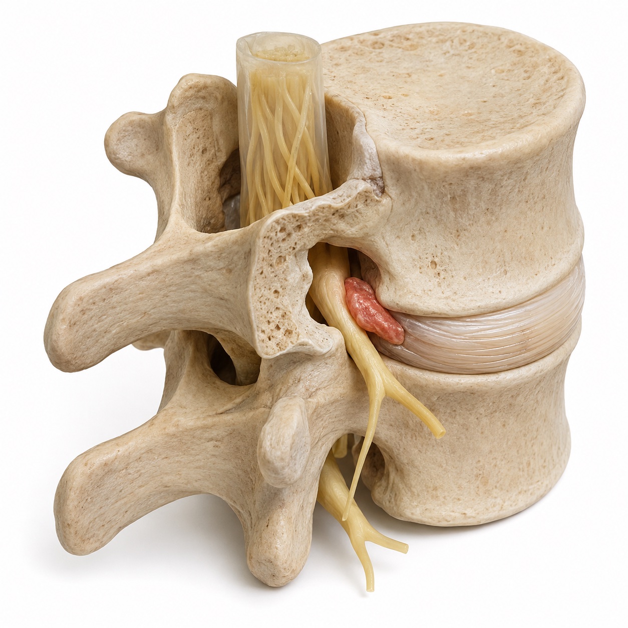
A nerve is still under pressure
A nerve may remain compressed after an operation, or a disc can herniate again. The location of the pressure needs to match the symptoms.
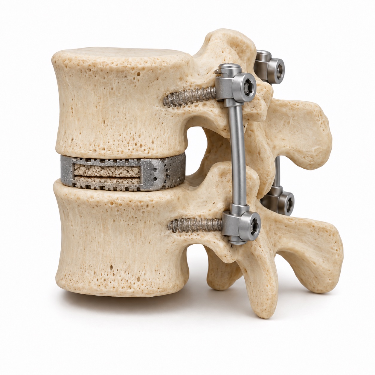
The bones have not joined
A non-union, also called pseudarthrosis, means the intended fusion has not formed a solid bone connection. Hardware alone does not prove that the bone has healed.
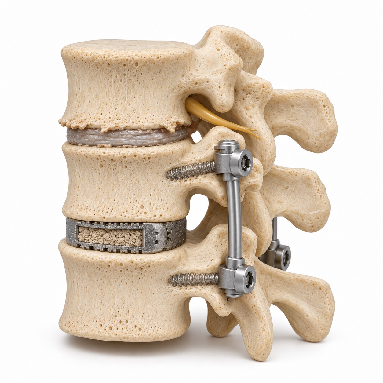
A nearby level has worn down
The earlier surgery may have worked well. Years later, wear at the level above or below it can become a new source of symptoms.
Simplified lumbar teaching models, not patient scans. Revision also involves the neck; anatomy and treatment vary. Symptoms, examination, and imaging guide the decision.
More about the reasons for revision surgery
The word revision covers three very different situations. In the first, the original problem was never fully addressed: a disc fragment was missed, a nerve tunnel was not opened enough, or the wrong level was treated. In the second, the operation was done well but the fusion did not heal, which is called a non-union or pseudarthrosis. In the third, the operation worked, and years later the level next to it wore out from carrying extra load, which is called adjacent segment disease.
Each of these has a different fix, a different timeline, and a different likelihood of success. Sorting out which one you have is most of the work, and it cannot be done from symptoms alone. It needs the report from the first operation, the imaging from before and after it, and new imaging designed to answer specific questions.
Other reasons for revision include hardware that has loosened or broken, infection, a recurrent disc herniation at the same level, and the spine drifting out of alignment after a long fusion. These are less common but follow the same logic.
Who needs revision surgery, and who does not?
The single most useful clue is when the pain came back. Pain that never left after the first operation points to a problem that was not addressed, such as a missed fragment or an incompletely opened nerve tunnel. Pain that returned within weeks to a few months, often suddenly, suggests a recurrent disc herniation or an early hardware problem. Pain that came on gradually over months, with neck or back pain rather than arm or leg pain, and hardware that looks loose on X-ray, suggests a fusion that has not healed. Pain that returned after years of feeling well, often with symptoms at a slightly different level or in a different distribution, suggests adjacent segment disease.
Dr. Boody will ask you to reconstruct that timeline carefully, because it narrows the search before any new test is ordered.
Who does not need revision surgery? Anyone whose imaging shows nothing that explains the pain. Scar tissue around a nerve is normal after surgery and is rarely a reason to operate. A fusion that has not healed on X-ray but is not causing symptoms is usually watched. Dr. Boody co-authored a study of patients without radiographic union two years after single-level ACDF, and many of them did well over longer follow-up without another operation. Absence of fusion is a finding to interpret, not a diagnosis by itself.
What does the workup involve?
Before another operation
What does each part of the workup tell us?
What was done before?
The operative note identifies the levels treated and the implants used. Comparing old and new images helps show what has changed.
Bring the prior surgery report and imaging if you have them. The team can help request missing records.
Has the fusion healed?
CT looks at the bone bridge, screws, and cage. Bending X-rays can help assess movement at the treated or neighboring levels.
These tests answer different questions. Dr. Boody chooses them based on the history and the existing images.
What explains the symptoms?
MRI helps evaluate the nerves and surrounding tissue. A targeted injection may help test whether a suspected area is contributing to the pain.
The examination, symptom pattern, and imaging need to fit together before a surgical plan is made.
Not everyone needs every test. The workup is chosen to answer the questions raised by your previous surgery and current symptoms.
Read the full workup, including injections and bone health checks
- The operative note. It records what was done, at which levels, with which implants. Without it, the surgeon is guessing at the geometry of the prior surgery. Your previous surgeon's office can send it; the team will help you request it.
- Old imaging. The MRI from before the first operation and any X-rays since. Comparing then and now is often more informative than any single new scan.
- CT scan. The best test for whether a fusion has healed and whether screws are loose or a cage has shifted. Thin cuts show bridging bone or the lack of it.
- Flexion and extension X-rays. Standing X-rays bending forward and back. Abnormal motion at a fused level means the fusion has not healed; abnormal motion at the next level suggests adjacent segment instability.
- MRI, often with contrast. Contrast helps distinguish scar tissue, which enhances, from a recurrent disc fragment, which does not. When hardware makes MRI hard to read, a CT myelogram, with dye injected around the nerves, is used instead.
- Diagnostic injection. A numbing injection at a specific nerve root, facet joint, or the sacroiliac joint can confirm or rule out a pain source before committing to surgery at that spot. Temporary relief that matches the injection is strong evidence.
- Blood tests and bone density. Inflammatory markers if infection is a possibility, blood sugar control, and a bone density test when a fusion is planned in someone with weak bone.
What happens during the operation?
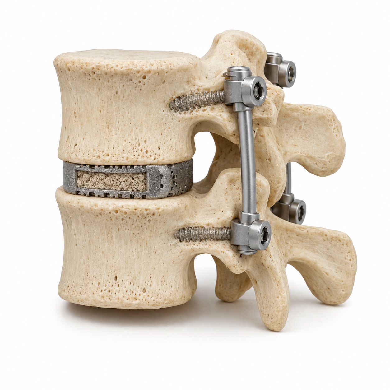
Keep what is working.
Stable hardware that is not causing trouble can often remain.
Repair what needs support.
Loose or broken implants may be exchanged. A symptomatic non-union may need additional graft and stabilization.
Address a neighboring level.
If a nearby level needs fusion, the construct may be extended. In selected cases, decompression alone is enough.
Walk through the operation, including scar tissue and hardware revision
- Anesthesia and positioning. General anesthesia with nerve monitoring in most revision cases. Positioning face down for posterior surgery or on the back for anterior neck surgery, matched to the plan.
- Incision. Usually through or alongside the old scar. In the neck, a revision from the front may deliberately use the opposite side to avoid scar, after checking that both vocal cords work normally. In the low back, a minimally invasive corridor away from the old midline scar is sometimes possible, and is described on the minimally invasive surgery page.
- Working through scar. Scar does not have the natural planes of unoperated tissue, and the nerve covering can be stuck to it. This part of a revision is slower and is where most of the extra risk lives.
- Hardware removal or exchange. Old screws and rods or a plate are removed if they are loose, broken, in the way, or irritating a structure. Larger or longer screws may be placed in the same holes, or new anchor points are chosen. Hardware that is solidly fused in and causing no trouble is often left alone.
- Rebuilding a non-union. For a neck fusion that did not heal, the common fix is to add stability from the back, with screws and rods and bone graft across the back of the level, rather than reopening the front. For a lumbar non-union, the cage may be exchanged and screws revised, with bone graft added.
- Extending a fusion. For adjacent segment disease, the worn level is decompressed and the existing construct is extended by one level, connecting to the old hardware where possible.
A one-level revision decompression may take one to two hours with a same-day or one-night stay. A revision fusion with hardware exchange typically takes three to five hours with a stay of two to four nights.
What are the alternatives?
Non-surgical care. Therapy, medication, and activity modification are tried again, often for longer than before a first operation, unless there is progressive weakness or spinal cord compression.
Injections. Both diagnostic and therapeutic. An injection that gives lasting relief can be repeated within limits and may make surgery unnecessary.
A different operation than you expected. Sometimes the best plan for a failed ACDF is a small posterior stabilization rather than redoing the front. Sometimes a level that wore out next to a fusion can be decompressed without extending the fusion. The alternative is often a smaller operation than the one you were told you would need.
What are the risks?
- Dural tear. The membrane around the nerves is often stuck to scar and can tear when it is freed. Studies report this in a substantial minority of lumbar revisions, well above the rate in first operations. It is repaired or sealed during surgery and may mean a day or two lying flat; it rarely has lasting consequences.
- Nerve injury. Nerves do not move aside as easily when scarred. Temporary new numbness or weakness is more common than after a first operation; permanent injury remains uncommon.
- Infection. Higher with longer operations, more blood loss, and previous hardware. Dr. Boody co-authored a consensus series on surgical site infection in spine surgery, and the prevention steps from that work are built into the plan.
- Blood loss and transfusion. Scar bleeds more. Larger revisions may require a transfusion, and blood-saving techniques are used routinely.
- Non-union again. Bone that failed to heal once has a higher chance of failing again, especially in smokers, in poorly controlled diabetes, and with weak bone. The plan addresses each of those before surgery.
- Specific to the neck. A second operation from the front carries a higher risk of voice change and swallowing difficulty, which is why the opposite side or a posterior approach is often chosen.
- Persistent pain. The most common disappointment after revision is not a complication but a result that falls short. This is discussed honestly below.
What is recovery like?
Recovery can resemble a first operation, with extra time for healing.
A longer recovery, with follow-up as the fusion matures over six to twelve months.
- First week
Start walking and protect the repair.
Short walks begin early. Soreness and lifting limits are expected; a brace or collar may be part of the plan.
- Weeks 2 to 6
Build a steady routine.
The wound is checked and walking increases. Return to desk work generally takes longer after fusion than after decompression.
- Months 2 to 3
Rebuild strength gradually.
Therapy focuses on strength and movement. Fusion patients continue lifting restrictions while imaging checks progress.
- Months 3 to 12
Let confirmed healing guide activity.
Revision fusions are followed closely. Heavier lifting and impact wait for clearance based on healing.
More about activity limits and recovery timing
First week
Walking from the first day. More incision and muscle soreness than the first time. A drain for a day or two. A brace or collar is more likely after a revision fusion, for comfort and to protect the construct. Lifting limited to about ten pounds.
Weeks 2 to 6
Wound check. Desk work at two to four weeks for decompression, four to eight weeks for fusion. Driving once off narcotic medication. Walking distance builds up. Nerve symptoms that were present for a long time before surgery improve slowly.
Months 2 to 3
Physical therapy focused on core strength and mechanics. Return to physical work for decompression patients. Fusion patients continue lifting limits. Follow-up X-rays check hardware and early fusion.
Months 3 to 12
A revision fusion is watched more closely and for longer, with X-rays or CT at intervals through the first year. Heavy lifting and impact are cleared as fusion is confirmed, typically between four and nine months. Long-term follow-up continues because the levels next to a longer construct carry more load.
What results can you expect?
The outcome tracks the reason. A missed disc fragment or an inadequately opened nerve tunnel, once corrected, often produces relief similar to a first operation. Adjacent segment disease that is decompressed and stabilized has good outcomes in published series, though the level beyond the new fusion now takes on the same extra load. Non-union repair improves pain for most patients whose symptoms genuinely came from the non-union, which is why the workup sometimes includes an injection to make sure they did.
Two of Dr. Boody's own studies inform how he sets expectations. His research on patients without radiographic fusion two years after ACDF showed that a non-union on X-ray does not automatically mean a bad outcome, so he does not recommend a revision for the X-ray alone. And his work on adjacent segment disease, including the ten-year adjacent-level outcomes after cervical disc replacement and fusion and a study on whether restoring segmental alignment in one-level fusions reduces adjacent segment disease, informs how he plans the new construct so the next level is protected as well as it can be.
What he will not do is promise that a revision will make you feel the way you did before your first operation. Some patients get there. The realistic goal is a specific improvement in a specific symptom, agreed on before surgery, with the risks stated in numbers.