


What is ACDF?
The same level, before and after ACDF
Look at the lower disc space in these matching views. The operation removes the source of pressure and supports the space left behind.
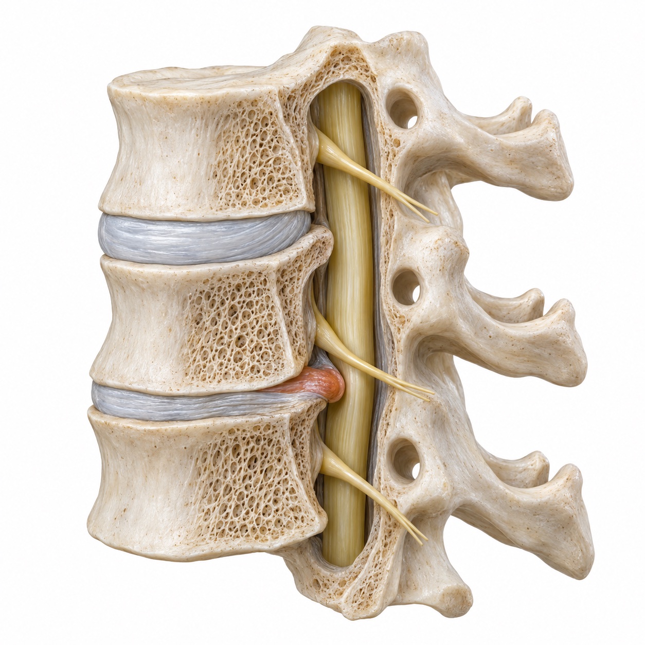
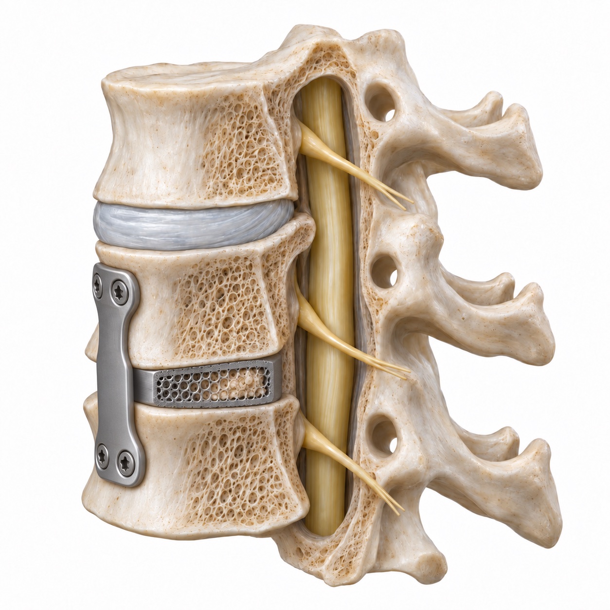
Educational illustrations of a one-level operation, not patient scans. Implant designs vary; the bone fusion is not immediate.
The name spells out the steps. Anterior means the surgeon reaches the spine from the front. Cervical means the neck. Discectomy means the disc is removed. Fusion means the two bones that used to sit on either side of that disc are joined with a bone graft so that, over a few months, they heal into one.
Why the front of the neck? Almost everything that pinches a nerve in the neck sits in front of it: the disc, the bone spurs that grow at the disc edges, and sometimes a thickened ligament. Coming from the front puts those structures directly in view. The approach passes between the windpipe and swallowing tube on one side and the large neck vessels on the other, through a natural plane that needs very little cutting. That is also why the neck muscles at the back are untouched and why most people are up and walking the same day.
Once the disc is out, the empty space is held open with a spacer, called a cage, made of a plastic polymer, titanium, or a block of donor bone. The cage is packed with bone graft. A small titanium plate is usually screwed across the front of the two vertebrae to hold everything still while the bone heals. Some cage designs have their own screws built in and do not need a separate plate.
Who is ACDF for, and who is it not for?
The two conditions that most often lead to ACDF are cervical radiculopathy, a pinched nerve root that sends pain, tingling, or weakness down a specific path in the arm, and cervical myelopathy, pressure on the spinal cord itself that shows up as clumsy hands, an unsteady walk, or changes in balance.
For radiculopathy, surgery is usually offered when symptoms have lasted more than six to twelve weeks despite medication, therapy, and sometimes an injection, or sooner if there is meaningful weakness. Most pinched nerves in the neck improve without surgery, so the first visit is often about confirming the diagnosis and buying time.
For myelopathy the calculation is different. The spinal cord does not tolerate pressure well over the long term, and a cord that is already showing signs of injury tends to lose function gradually rather than recover. Surgery is recommended earlier, to stop the decline, and ACDF is one of the main ways to do it when the compression comes from the front.
ACDF is a good fit when the level has already lost most of its motion, when there is arthritis in the small joints at the back of that level, when there is instability, or when more than one thing needs fixing at the same level. It is a less obvious choice when the level is still healthy and mobile apart from a single soft herniation. In that setting cervical disc replacement may preserve motion and is worth a direct conversation.
What happens during the operation?
Follow the operation
See what changes at each step.
Find what is pressing on the nerve or cord.
The MRI and examination identify the level that matches your symptoms. This example shows a herniated disc; bone spurs can also cause pressure.
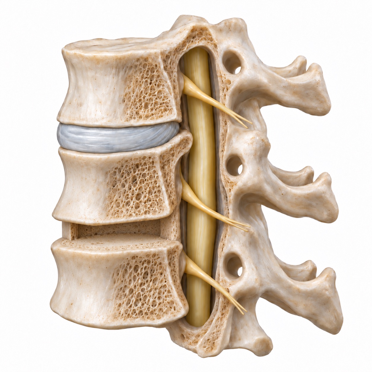
Remove the disc and free the nerves.
Through the front of the neck, the surgeon removes the damaged disc and any bone spurs causing pressure. The nerve openings are cleared as needed.
Place the graft and stabilize the level.
A cage containing bone graft fills the disc space. A plate and screws usually hold the two vertebrae steady while the graft heals into a solid bridge.
Simplified educational views. The approach and implant construct are chosen for your anatomy.
Read the full surgical steps
- Anesthesia. You are fully asleep with a breathing tube. An anesthesiologist monitors you throughout. For myelopathy cases, nerve monitoring leads are often placed on the arms and legs so the team can watch spinal cord signals during the operation.
- Incision. A horizontal cut about two inches long is made in a natural skin crease on the front of the neck, usually on the left side. Once healed it is hard to find.
- Reaching the spine. The thin muscle under the skin is opened, and the surgeon moves gently between the windpipe and swallowing tube on the inside and the carotid artery and jugular vein on the outside. No major muscle is cut. An X-ray confirms the correct level.
- Removing the disc. Under magnification, the disc is removed piece by piece, along with the bone spurs at its edges. The ligament behind the disc is opened if the herniation has pushed through it. The surgeon then widens the small tunnel where the nerve root exits so nothing presses on it.
- Placing the graft. The empty disc space is measured, and a cage of the right height is packed with bone graft and tapped into place. This restores the height the disc had lost and opens the nerve tunnels further.
- The plate. A titanium plate is set across the front of the two vertebrae and secured with four short screws. It acts as an internal splint while the bone heals. A final X-ray checks position.
A single level usually takes one to two hours of operating time. Two levels add roughly forty-five minutes to an hour. Most one-level and many two-level patients go home the same day or after a single night; a stay of one to two nights is more likely with additional levels, with myelopathy, or with other medical conditions.
What are the alternatives?
Cervical disc replacement. The same approach from the front and the same disc removal, but instead of a fused level the disc is replaced with an implant that keeps the level moving. It is the main motion-preserving alternative for one or two levels when the level is still healthy enough to move well. The comparison below covers the trade-offs.
Posterior foraminotomy. A small opening made from the back of the neck can free a nerve root when the herniation sits out to the side. It avoids fusion and hardware, but it does not address pressure in the middle of the canal, and it does not stabilize the level.
Posterior decompression for myelopathy. When the cord is compressed over several levels, or from behind, an operation from the back (laminoplasty or laminectomy with fusion) may be a better fit than a long fusion from the front.
Fusion or disc replacement: see the difference
Both remove the damaged disc. The difference is what replaces it and whether that level is intended to move.
ACDF
Goal: one solid, stable level- Fuses the level; motion at that level is given up
- Works for arthritic, collapsed, or unstable levels
- Handles bone spurs, instability, and multi-level disease
- Long track record; fusion rates for one level are high
- Small risk of non-union; neighboring levels take on more load over time
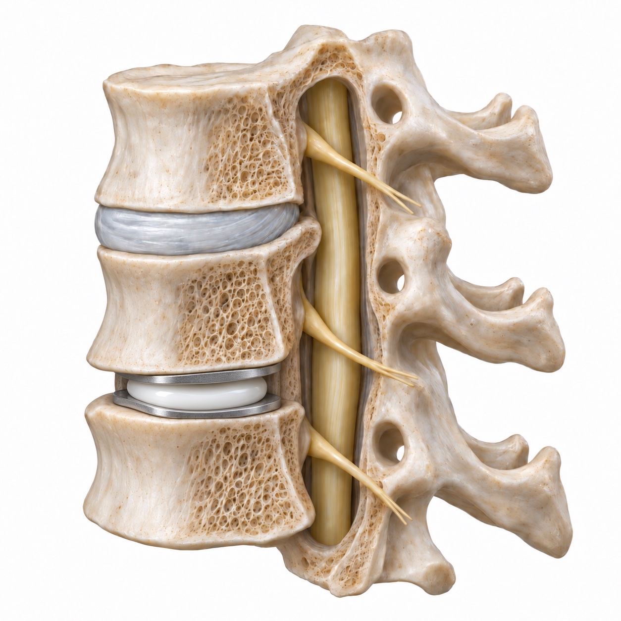
Cervical disc replacement
Goal: preserve motion at the level- Keeps the level moving
- Needs a healthy, mobile level without significant facet arthritis or instability
- Usually one or two levels of soft disc disease
- Long-term trial data now reach twenty years
- Bone can form around the implant over time (heterotopic ossification); not everyone qualifies
Generic implant illustrations. Your scans, joint health, and spinal stability help determine which option fits.
What are the risks?
- Swallowing soreness. Nearly everyone has some difficulty swallowing in the first days because the swallowing tube was moved aside. For most it fades within two to six weeks. A small percentage of patients report some lingering sensation beyond three months, more often after multi-level surgery.
- Hoarseness. The nerve that moves one vocal cord runs through the surgical field. Temporary hoarseness occurs in a minority of patients and almost always resolves; permanent voice change is rare.
- Non-union (pseudarthrosis). The bone graft does not always heal into solid bone. Studies report non-union in roughly 5 to 10 percent of single-level fusions, and the rate rises with each additional level, with smoking, and with poorly controlled diabetes. Non-union does not automatically mean another operation; see the results section below.
- Adjacent-level disease. Once a level is fused, the levels above and below move a little more to compensate. Over years, some of them wear faster. Studies report that roughly 2 to 3 percent of ACDF patients per year develop symptoms at a neighboring level, which adds up to about one in four over ten years. Only a fraction of those need surgery.
- Hardware problems. Screws can loosen or a plate can shift, especially if the fusion is slow to heal. Rarely a plate irritates the swallowing tube and needs removal after the fusion has healed.
- Nerve or spinal cord injury. Rare, but the most serious risk. Nerve monitoring and magnification lower it. A temporary weakness of the shoulder (C5 palsy) occurs in a small number of patients after decompression and usually recovers over months.
- Bleeding in the neck. Uncommon, but a collection of blood in the neck after surgery can press on the airway, which is one reason you are watched closely for the first hours and told what to look for at home.
- Infection, blood clots, anesthesia risks. All low with the anterior approach, and lower still with good preparation: stopping smoking, controlling blood sugar, and walking early.
What is recovery like?
-
First week
Days 1 to 7
- Sore throat and a stiff neck. Soft foods help. A soft collar if it makes you comfortable.
- Walk several times a day. No lifting over about ten pounds.
- Most people need pain medication for a few days, then mostly over-the-counter options.
-
Weeks 2 to 6
Early healing
- Swallowing settles. Driving once you are off narcotic pain medication and can turn your head comfortably.
- Desk work usually resumes in this window. Gentle walking builds up; no overhead lifting or impact yet.
- Follow-up X-ray to confirm position.
-
Months 2 to 3
Fusion forming
- Physical therapy for posture and neck mechanics if needed.
- Return to physical jobs and to most exercise, usually with a gradual ramp.
- Arm symptoms have typically improved by now; numbness can be the slowest to fade.
-
Months 3 to 12
Longer-term recovery
- The bone graft matures into solid fusion, confirmed on X-ray or CT between three and twelve months.
- Full return to sport and heavy work is cleared once fusion is progressing.
- Long-term follow-up watches the neighboring levels.
What results can you expect?
Arm pain usually improves quickly, often within days. Weakness recovers more slowly, over weeks to months, and numbness is the least predictable symptom. Neck pain itself is the symptom ACDF fixes least reliably, which is why Dr. Boody is careful to separate the symptom you want gone from the one the operation is designed to treat.
Fusion rates for a one-level ACDF with a plate are high, and most published series report that the large majority of levels are solidly healed within a year. But an X-ray that fails to show union is not the whole story. Dr. Boody co-authored a study in Global Spine Journal (2020) following patients who had no radiographic sign of fusion two years after a single-level ACDF. Many of those patients did well over longer follow-up without further surgery. The practical lesson is that a non-union is a finding to be interpreted alongside your symptoms, not an automatic reason to operate again. When it does hurt, the fix is usually a small operation from the back of the neck to add stability, described on the revision surgery page.
Over decades, the question shifts from the fused level to its neighbors. Adjacent-level disease is the main long-term trade-off of any fusion, and it is the reason motion-preserving surgery exists. Whether restoring the natural curve at the fused level protects the neighboring ones is an open question that Dr. Boody has studied directly, in a 2020 paper on segmental lordosis in one-level fusions. Regardless of the answer, the right plan is a periodic check-in rather than a set of new X-rays every year.